 Movie
Movie Controller

[English] 日本語
 Yorodumi
- PDB-5otu: Extracellular domain of GLP-1 receptor in complex with GLP-1 vari... -
+ Open data

- Basic information
| Entry | Database: PDB / ID: 5otu | ||||||
|---|---|---|---|---|---|---|---|
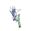
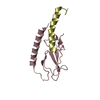
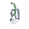
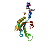
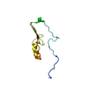
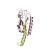
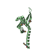
| Title | Extracellular domain of GLP-1 receptor in complex with GLP-1 variant Ala8Hcs/Thr11Hcs | ||||||
 Components Components |
| ||||||
 Keywords Keywords | SIGNALING PROTEIN / glucagon-like peptide 1 / GPCR / cyclic peptides | ||||||
| Function / homology |  Function and homology information Function and homology informationglucagon receptor binding / glucagon-like peptide 1 receptor activity / glucagon receptor activity / : / positive regulation of blood pressure / hormone secretion / post-translational protein targeting to membrane, translocation / feeding behavior / negative regulation of execution phase of apoptosis / response to psychosocial stress ...glucagon receptor binding / glucagon-like peptide 1 receptor activity / glucagon receptor activity / : / positive regulation of blood pressure / hormone secretion / post-translational protein targeting to membrane, translocation / feeding behavior / negative regulation of execution phase of apoptosis / response to psychosocial stress / regulation of heart contraction / positive regulation of calcium ion import / regulation of insulin secretion / peptide hormone binding / Synthesis, secretion, and deacylation of Ghrelin / activation of adenylate cyclase activity / negative regulation of blood pressure / cellular response to glucagon stimulus / positive regulation of gluconeogenesis / positive regulation of insulin secretion involved in cellular response to glucose stimulus / response to activity / gluconeogenesis / hormone activity / adenylate cyclase-modulating G protein-coupled receptor signaling pathway / positive regulation of insulin secretion / Glucagon signaling in metabolic regulation / Synthesis, secretion, and inactivation of Glucagon-like Peptide-1 (GLP-1) / transmembrane signaling receptor activity / Glucagon-type ligand receptors / Glucagon-like Peptide-1 (GLP1) regulates insulin secretion / glucose homeostasis / adenylate cyclase-activating G protein-coupled receptor signaling pathway / positive regulation of cytosolic calcium ion concentration / secretory granule lumen / G alpha (s) signalling events / G alpha (q) signalling events / learning or memory / positive regulation of ERK1 and ERK2 cascade / cell surface receptor signaling pathway / endoplasmic reticulum lumen / G protein-coupled receptor signaling pathway / receptor ligand activity / signaling receptor binding / negative regulation of apoptotic process / : / extracellular region / membrane / identical protein binding / plasma membrane Similarity search - Function | ||||||
| Biological species |  Homo sapiens (human) Homo sapiens (human) | ||||||
| Method |  X-RAY DIFFRACTION / SYNCHROTRON / MOLECULAR REPLACEMENT / Resolution: 1.8 Å X-RAY DIFFRACTION / SYNCHROTRON / MOLECULAR REPLACEMENT / Resolution: 1.8 Å | ||||||
 Authors Authors | Mortensen, S. | ||||||
 Citation Citation | Journal: Biochemistry / Year: 2018 Title: alpha-Helix or beta-Turn? An Investigation into N-Terminally Constrained Analogues of Glucagon-like Peptide 1 (GLP-1) and Exendin-4. Authors: Oddo, A. / Mortensen, S. / Thogersen, H. / De Maria, L. / Hennen, S. / McGuire, J.N. / Kofoed, J. / Linderoth, L. / Reedtz-Runge, S. | ||||||
| History |
|
- Structure visualization
| Structure viewer | Molecule: MolmilJmol/JSmol |
|---|
- Downloads & links
-Download
| PDBx/mmCIF format | 5otu.cif.gz | 123.4 KB | Display | PDBx/mmCIF format |
|---|---|---|---|---|
| PDB format | pdb5otu.ent.gz | 95.4 KB | Display | PDB format |
| PDBx/mmJSON format | 5otu.json.gz | Tree view | PDBx/mmJSON format | |
| Others |  Other downloads Other downloads |
-Validation report
| Arichive directory | https://data.pdbj.org/pub/pdb/validation_reports/ot/5otuftp://data.pdbj.org/pub/pdb/validation_reports/ot/5otu | HTTPS FTP |
|---|
-Related structure data
| Related structure data |  5ottC  5otvC  5otwC  5otxC  4zgmS S: Starting model for refinement C: citing same article ( |
|---|---|
| Similar structure data |


-Links
 PDBj
PDBj


- Assembly
| Deposited unit | 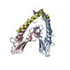
| ||||||||
|---|---|---|---|---|---|---|---|---|---|
| 1 | 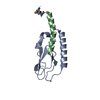
| ||||||||
| 2 | 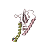
| ||||||||
| Unit cell |
|
-Components
| #1: Protein | Mass: 13547.892 Da / Num. of mol.: 2 / Fragment: extracellular domain, UNP residues 24-139 Source method: isolated from a genetically manipulated source Source: (gene. exp.) Homo sapiens (human) / Gene: GLP1R / Production host:  #2: Protein/peptide | Mass: 3421.857 Da / Num. of mol.: 2 / Mutation: A8HCS, T11HCS / Source method: obtained synthetically / Source: (synth.) Homo sapiens (human) / References: UniProt: P01275#3: Water | ChemComp-HOH / |  Mass: 18.015 Da / Num. of mol.: 212 / Source method: isolated from a natural source / Formula: H2O Mass: 18.015 Da / Num. of mol.: 212 / Source method: isolated from a natural source / Formula: H2OHas protein modification | Y | |
|---|
-Experimental details
-Experiment
| Experiment | Method: X-RAY DIFFRACTION / Number of used crystals: 1 |
|---|
- Sample preparation
| Crystal | Density Matthews: 2.02 Å3/Da / Density % sol: 39.18 % |
|---|---|
| Crystal grow | Temperature: 293 K / Method: vapor diffusion, sitting drop Details: Morpheus from Molecular Dimensions, solution G1: 0.1 M Carboxylic acids, 0.1 Buffer system 1 pH 6.5, 20% (v/v) PEG500mme, 10% (w/v) PEG20000 |
-Data collection
| Diffraction | Mean temperature: 100 K |
|---|---|
| Diffraction source | Source: SYNCHROTRON / Site: SLS  / Beamline: X06DA / Wavelength: 1 Å / Beamline: X06DA / Wavelength: 1 Å |
| Detector | Type: DECTRIS PILATUS 2M / Detector: PIXEL / Date: Feb 4, 2017 |
| Radiation | Protocol: SINGLE WAVELENGTH / Monochromatic (M) / Laue (L): M / Scattering type: x-ray |
| Radiation wavelength | Wavelength: 1 Å / Relative weight: 1 |
| Reflection | Resolution: 1.8→42.535 Å / Num. obs: 24985 / % possible obs: 99.7 % / Redundancy: 3.4 % / CC1/2: 0.999 / Rmerge(I) obs: 0.045 / Rpim(I) all: 0.029 / Rsym value: 0.054 / Net I/σ(I): 15.6 |
| Reflection shell | Resolution: 1.8→1.84 Å / Redundancy: 3.53 % / Rmerge(I) obs: 0.482 / Mean I/σ(I) obs: 2.7 / Num. measured obs: 5444 / Num. unique all: 1544 / CC1/2: 0.851 / Rpim(I) all: 0.298 / Rrim(I) all: 0.568 / % possible all: 99.68 |
- Processing
| Software |
| |||||||||||||||||||||||||||||||||||||||||||||||||||||||||||||||||||||||||||||||||||||||||||||||||||||||||||||||||||||||||||||
|---|---|---|---|---|---|---|---|---|---|---|---|---|---|---|---|---|---|---|---|---|---|---|---|---|---|---|---|---|---|---|---|---|---|---|---|---|---|---|---|---|---|---|---|---|---|---|---|---|---|---|---|---|---|---|---|---|---|---|---|---|---|---|---|---|---|---|---|---|---|---|---|---|---|---|---|---|---|---|---|---|---|---|---|---|---|---|---|---|---|---|---|---|---|---|---|---|---|---|---|---|---|---|---|---|---|---|---|---|---|---|---|---|---|---|---|---|---|---|---|---|---|---|---|---|---|---|
| Refinement | Method to determine structure: MOLECULAR REPLACEMENT Starting model: 4ZGM Resolution: 1.8→42.535 Å / SU ML: 0.18 / Cross valid method: FREE R-VALUE / σ(F): 1.36 / Phase error: 22.58
| |||||||||||||||||||||||||||||||||||||||||||||||||||||||||||||||||||||||||||||||||||||||||||||||||||||||||||||||||||||||||||||
| Solvent computation | Shrinkage radii: 0.9 Å / VDW probe radii: 1.11 Å | |||||||||||||||||||||||||||||||||||||||||||||||||||||||||||||||||||||||||||||||||||||||||||||||||||||||||||||||||||||||||||||
| Refinement step | Cycle: LAST / Resolution: 1.8→42.535 Å
| |||||||||||||||||||||||||||||||||||||||||||||||||||||||||||||||||||||||||||||||||||||||||||||||||||||||||||||||||||||||||||||
| Refine LS restraints |
| |||||||||||||||||||||||||||||||||||||||||||||||||||||||||||||||||||||||||||||||||||||||||||||||||||||||||||||||||||||||||||||
| LS refinement shell |
| |||||||||||||||||||||||||||||||||||||||||||||||||||||||||||||||||||||||||||||||||||||||||||||||||||||||||||||||||||||||||||||
| Refinement TLS params. | Method: refined / Refine-ID: X-RAY DIFFRACTION
| |||||||||||||||||||||||||||||||||||||||||||||||||||||||||||||||||||||||||||||||||||||||||||||||||||||||||||||||||||||||||||||
| Refinement TLS group |
|
