- PDB-3ue2: Crystal structure of a RNA binding domain of poly-U binding splic... -
+
Open data
ID or keywords:
Loading...
-
Basic information
Entry
Database: PDB / ID: 3ue2
Title
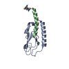
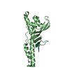
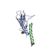
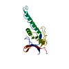
Crystal structure of a RNA binding domain of poly-U binding splicing factor 60KDa (PUF60) from Homo sapiens at 1.23 A resolution
Components
Poly(U)-binding-splicing factor PUF60
Keywords
RNA BINDING PROTEIN / RNA RECOGNITION MOTIF / RRM / RNA BINDING DOMAIN / SPLICING / Structural Genomics / Joint Center for Structural Genomics / JCSG / Protein Structure Initiative / PSI-BIOLOGY / Partnership for T-Cell Biology / TCELL
Function / homology
Function and homology information
mRNA splice site recognition / alternative mRNA splicing, via spliceosome / regulation of alternative mRNA splicing, via spliceosome / mRNA Splicing - Major Pathway / cell junction / cadherin binding / ribonucleoprotein complex / apoptotic process / DNA binding / RNA binding ...mRNA splice site recognition / alternative mRNA splicing, via spliceosome / regulation of alternative mRNA splicing, via spliceosome / mRNA Splicing - Major Pathway / cell junction / cadherin binding / ribonucleoprotein complex / apoptotic process / DNA binding / RNA binding / nucleoplasm / identical protein binding Similarity search - Function
Mass: 18.015 Da / Num. of mol.: 189 / Source method: isolated from a natural source / Formula: H2O
Has protein modification
Y
Sequence details
THIS CONSTRUCT (RESIDUES 443-559) WAS EXPRESSED WITH A PURIFICATION TAG MGSDKIHHHHHHENLYFQG. THE ...THIS CONSTRUCT (RESIDUES 443-559) WAS EXPRESSED WITH A PURIFICATION TAG MGSDKIHHHHHHENLYFQG. THE TAG WAS REMOVED WITH TEV PROTEASE LEAVING ONLY A GLYCINE (0) FOLLOWED BY THE TARGET SEQUENCE. RESIDUE NUMBERING IS BASED ON ISOFORM 1 OF UNIPROTKB Q9UHX1.
-
Experimental details
-
Experiment
Experiment
Method: X-RAY DIFFRACTION / Number of used crystals: 1
-
Sample preparation
Crystal
Density Matthews: 1.87 Å3/Da / Density % sol: 34.1 %
Crystal grow
Temperature: 277 K / Method: vapor diffusion, sitting drop / pH: 6 Details: 2.4 M ammonium sulfate, 0.1M MES pH 6.0, NANODROP, VAPOR DIFFUSION, SITTING DROP, temperature 277K
Resolution: 1.23→29.553 Å / Num. all: 30048 / Num. obs: 30048 / % possible obs: 99.8 % / Redundancy: 3.4 % / Rsym value: 0.08 / Net I/σ(I): 8.3
Reflection shell
Diffraction-ID: 1
Resolution (Å)
Redundancy (%)
Rmerge(I) obs
Mean I/σ(I) obs
Num. measured all
Num. unique all
Rsym value
% possible all
1.23-1.26
3.4
0.612
1.3
7515
2198
0.612
100
1.26-1.3
3.4
0.508
1.5
7240
2119
0.508
100
1.3-1.33
3.4
0.437
1.7
7120
2078
0.437
100
1.33-1.38
3.4
0.345
2.2
6974
2039
0.345
100
1.38-1.42
3.5
0.303
2.5
6702
1940
0.303
99.9
1.42-1.47
3.4
0.24
3.2
6623
1929
0.24
100
1.47-1.53
3.4
0.198
3.3
6260
1825
0.198
100
1.53-1.59
3.4
0.156
4.7
6094
1771
0.156
100
1.59-1.66
3.4
0.13
5.7
5881
1711
0.13
100
1.66-1.74
3.4
0.11
6.6
5594
1633
0.11
99.9
1.74-1.83
3.4
0.09
8
5308
1552
0.09
99.9
1.83-1.94
3.4
0.073
9.7
4951
1455
0.073
99.8
1.94-2.08
3.4
0.065
10.3
4740
1400
0.065
99.8
2.08-2.25
3.3
0.067
9.9
4286
1281
0.067
99.5
2.25-2.46
3.3
0.067
9.6
3924
1200
0.067
99.5
2.46-2.75
3.1
0.064
9.9
3383
1081
0.064
99.1
2.75-3.18
3.2
0.06
10.2
3093
964
0.06
99.1
3.18-3.89
3.5
0.042
14.9
2917
834
0.042
99.7
3.89-5.5
3.4
0.041
15.5
2278
662
0.041
99.3
5.5-29.553
3.1
0.051
12.1
1173
376
0.051
96.5
-
Phasing
Phasing
Method: MAD
-
Processing
Software
Name
Version
Classification
NB
MolProbity
3beta29
modelbuilding
PDB_EXTRACT
3.1
dataextraction
SOLVE
phasing
SCALA
3.3.20
datascaling
REFMAC
5.5.0110
refinement
MOSFLM
datareduction
Refinement
Method to determine structure: MAD / Resolution: 1.23→29.553 Å / Cor.coef. Fo:Fc: 0.977 / Cor.coef. Fo:Fc free: 0.97 / Occupancy max: 1 / Occupancy min: 0.06 / SU B: 1.52 / SU ML: 0.03 / Cross valid method: THROUGHOUT / σ(F): 0 / ESU R Free: 0.042 Stereochemistry target values: MAXIMUM LIKELIHOOD WITH PHASES Details: 1.HYDROGENS HAVE BEEN ADDED IN THE RIDING POSITIONS. 2.A MET-INHIBITION PROTOCOL WAS USED FOR SELENOMETHIONINE INCORPORATION DURING PROTEIN EXPRESSION. THE OCCUPANCY OF THE SE ATOMS IN THE ...Details: 1.HYDROGENS HAVE BEEN ADDED IN THE RIDING POSITIONS. 2.A MET-INHIBITION PROTOCOL WAS USED FOR SELENOMETHIONINE INCORPORATION DURING PROTEIN EXPRESSION. THE OCCUPANCY OF THE SE ATOMS IN THE MSE RESIDUES WAS REDUCED TO 0.75 FOR THE REDUCED SCATTERING POWER DUE TO PARTIAL S-MET INCORPORATION. 3.SULFATE (SO4) FROM THE CRYSTALLIZATION SOLUTION HAS BEEN MODELED IN THE SOLVENT STRUCTURE.
Rfactor
Num. reflection
% reflection
Selection details
Rfree
0.1601
1520
5.1 %
RANDOM
Rwork
0.1341
-
-
-
obs
0.1355
29999
99.66 %
-
Solvent computation
Ion probe radii: 0.8 Å / Shrinkage radii: 0.8 Å / VDW probe radii: 1.4 Å / Solvent model: BABINET MODEL WITH MASK
In the structure databanks used in Yorodumi, some data are registered as the other names, "COVID-19 virus" and "2019-nCoV". Here are the details of the virus and the list of structure data.
Jan 31, 2019. EMDB accession codes are about to change! (news from PDBe EMDB page)
EMDB accession codes are about to change! (news from PDBe EMDB page)
The allocation of 4 digits for EMDB accession codes will soon come to an end. Whilst these codes will remain in use, new EMDB accession codes will include an additional digit and will expand incrementally as the available range of codes is exhausted. The current 4-digit format prefixed with “EMD-” (i.e. EMD-XXXX) will advance to a 5-digit format (i.e. EMD-XXXXX), and so on. It is currently estimated that the 4-digit codes will be depleted around Spring 2019, at which point the 5-digit format will come into force.
The EM Navigator/Yorodumi systems omit the EMD- prefix.
Related info.:Q: What is EMD? / ID/Accession-code notation in Yorodumi/EM Navigator
Yorodumi is a browser for structure data from EMDB, PDB, SASBDB, etc.
This page is also the successor to EM Navigator detail page, and also detail information page/front-end page for Omokage search.
The word "yorodu" (or yorozu) is an old Japanese word meaning "ten thousand". "mi" (miru) is to see.
Related info.:EMDB / PDB / SASBDB / Comparison of 3 databanks / Yorodumi Search / Aug 31, 2016. New EM Navigator & Yorodumi / Yorodumi Papers / Jmol/JSmol / Function and homology information / Changes in new EM Navigator and Yorodumi
 Movie
Movie Controller

 Yorodumi
 Open data
 Basic information
 Components
 Keywords
 Function and homology information
 Homo sapiens (human)
 X-RAY DIFFRACTION /
  Authors
 Citation
 Structure visualization
 Downloads & links
 Other downloads
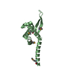


 PDBj
PDBj


 Assembly
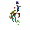


 Mass: 96.063 Da / Num. of mol.: 2 / Source method: obtained synthetically / Formula: SO4
 Mass: 18.015 Da / Num. of mol.: 189 / Source method: isolated from a natural source / Formula: H2O
 Sample preparation
 / Beamline: 8.2.2 / Wavelength: 0.9537,0.9796,0.9794
 Processing