 Movie
Movie Controller


+ Open data

- Basic information
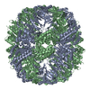
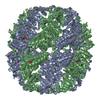
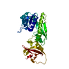
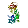
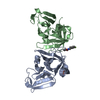
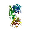
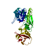
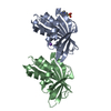
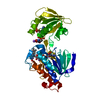
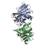
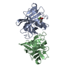
| Entry | Database: PDB / ID: 1e0r | ||||||
|---|---|---|---|---|---|---|---|
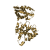
| Title | Beta-apical domain of thermosome | ||||||
 Components Components | THERMOSOME | ||||||
 Keywords Keywords | CHAPERONIN / HSP60 / THERMOSOME / TCP1 / GROEL / THERMOPLASMA ACIDOPHILUM | ||||||
| Function / homology |  Function and homology information Function and homology informationchaperonin ATPase / ATP-dependent protein folding chaperone / : / ATP hydrolysis activity / protein-containing complex / ATP binding / cytoplasm Similarity search - Function | ||||||
| Biological species |   THERMOPLASMA ACIDOPHILUM (acidophilic) THERMOPLASMA ACIDOPHILUM (acidophilic) | ||||||
| Method |  X-RAY DIFFRACTION / SYNCHROTRON / MOLECULAR REPLACEMENT / Resolution: 2.8 Å X-RAY DIFFRACTION / SYNCHROTRON / MOLECULAR REPLACEMENT / Resolution: 2.8 Å | ||||||
 Authors Authors | Bosch, G. / Baumeister, W. / Essen, L.-O. | ||||||
 Citation Citation | Journal: J.Mol.Biol. / Year: 2000 Title: Crystal Structure of the Beta-Apical Domain from Thermosome Reveals Structural Plasticity in Protrusion Region Authors: Bosch, G. / Baumeister, W. / Essen, L.-O. #1: Journal: Cell(Cambridge,Mass.) / Year: 1998Title: Crystal Structure of the Thermosome, the Archaeal Chaperonin and Homolog of Cct Authors: Ditzel, L. / Lowe, J. / Stock, D. / Stetter, K.O. / Huber, H. / Huber, R. / Steinbacher, S. #2: Journal: Cell(Cambridge,Mass.) / Year: 1997Title: Structure of the Substrate-Binding Domain of the Thermosome, an Archaeal Group II Chaperonin Authors: Klumpp, M. / Baumeister, W. / Essen, L.-O. | ||||||
| History |
|
- Structure visualization
| Structure viewer | Molecule: MolmilJmol/JSmol |
|---|
- Downloads & links
-Download
| PDBx/mmCIF format | 1e0r.cif.gz | 42 KB | Display | PDBx/mmCIF format |
|---|---|---|---|---|
| PDB format | pdb1e0r.ent.gz | 28.8 KB | Display | PDB format |
| PDBx/mmJSON format | 1e0r.json.gz | Tree view | PDBx/mmJSON format | |
| Others |  Other downloads Other downloads |
-Validation report
| Arichive directory | https://data.pdbj.org/pub/pdb/validation_reports/e0/1e0rftp://data.pdbj.org/pub/pdb/validation_reports/e0/1e0r | HTTPS FTP |
|---|
-Related structure data
| Related structure data |  1assS S: Starting model for refinement |
|---|---|
| Similar structure data |

-Links
 PDBj
PDBj
- Assembly
| Deposited unit | 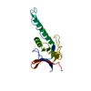
| ||||||||
|---|---|---|---|---|---|---|---|---|---|
| 1 | 
| ||||||||
| Unit cell |
|
-Components
| #1: Protein | Mass: 17763.393 Da / Num. of mol.: 1 / Fragment: SUBSTRATE-BINDING DOMAIN / Mutation: YES Source method: isolated from a genetically manipulated source Details: LYS 364 IS FOLLOWED BY ASN 365 AND HEXAHISTIDINE TAIL Source: (gene. exp.) THERMOPLASMA ACIDOPHILUM (acidophilic) / Cellular location: CYTOPLASM / Plasmid: PET22B / Cellular location (production host): CYTOPLASM / Gene (production host): THSB / Production host:  | ||
|---|---|---|---|
| Compound details | CHAIN B ENGINEERED| Sequence details | HIS: HISTIDINE TAG FROM HIS 368 - HIS 372 NOT SEEN IN ELECTRON DENSITY | |
-Experimental details
-Experiment
| Experiment | Method: X-RAY DIFFRACTION / Number of used crystals: 1 |
|---|
- Sample preparation
| Crystal | Density Matthews: 2.83 Å3/Da / Density % sol: 50 % | ||||||||||||||||||||||||||||||
|---|---|---|---|---|---|---|---|---|---|---|---|---|---|---|---|---|---|---|---|---|---|---|---|---|---|---|---|---|---|---|---|
| Crystal grow | pH: 9.1 / Details: pH 9.10 | ||||||||||||||||||||||||||||||
| Crystal grow | *PLUS Temperature: 20 ℃ / Method: vapor diffusion, hanging drop / pH: 9.1 | ||||||||||||||||||||||||||||||
| Components of the solutions | *PLUS
|
-Data collection
| Diffraction | Mean temperature: 100 K |
|---|---|
| Diffraction source | Source: SYNCHROTRON / Site: EMBL/DESY, HAMBURG  / Beamline: BW7A / Wavelength: 0.99 / Beamline: BW7A / Wavelength: 0.99 |
| Detector | Type: MARRESEARCH / Detector: IMAGE PLATE / Date: Jun 15, 1998 / Details: MIRROR |
| Radiation | Protocol: SINGLE WAVELENGTH / Monochromatic (M) / Laue (L): M / Scattering type: x-ray |
| Radiation wavelength | Wavelength: 0.99 Å / Relative weight: 1 |
| Reflection | Resolution: 2.8→25 Å / Num. obs: 5170 / % possible obs: 97.8 % / Observed criterion σ(I): 0 / Redundancy: 3.7 % / Biso Wilson estimate: 95 Å2 / Rmerge(I) obs: 0.041 / Net I/σ(I): 27 |
| Reflection shell | Resolution: 2.8→2.87 Å / Rmerge(I) obs: 0.393 / Mean I/σ(I) obs: 3.3 / % possible all: 96.8 |
| Reflection | *PLUS Num. measured all: 19163 |
| Reflection shell | *PLUS % possible obs: 96.8 % |
- Processing
| Software |
| ||||||||||||||||||||||||||||||||||||||||||||||||||||||||||||||||||||||||||||||||
|---|---|---|---|---|---|---|---|---|---|---|---|---|---|---|---|---|---|---|---|---|---|---|---|---|---|---|---|---|---|---|---|---|---|---|---|---|---|---|---|---|---|---|---|---|---|---|---|---|---|---|---|---|---|---|---|---|---|---|---|---|---|---|---|---|---|---|---|---|---|---|---|---|---|---|---|---|---|---|---|---|---|
| Refinement | Method to determine structure: MOLECULAR REPLACEMENT Starting model: PDB ENTRY 1ASS WITHOUT I245-K274 Resolution: 2.8→15 Å / Data cutoff high absF: 10000 / Isotropic thermal model: RESTRAINED / Cross valid method: THROUGHOUT / σ(F): 2
| ||||||||||||||||||||||||||||||||||||||||||||||||||||||||||||||||||||||||||||||||
| Solvent computation | Solvent model: FLAT MODEL / Bsol: 35.8 Å2 / ksol: 0.293 e/Å3 | ||||||||||||||||||||||||||||||||||||||||||||||||||||||||||||||||||||||||||||||||
| Displacement parameters | Biso mean: 76 Å2
| ||||||||||||||||||||||||||||||||||||||||||||||||||||||||||||||||||||||||||||||||
| Refinement step | Cycle: LAST / Resolution: 2.8→15 Å
| ||||||||||||||||||||||||||||||||||||||||||||||||||||||||||||||||||||||||||||||||
| Refine LS restraints |
| ||||||||||||||||||||||||||||||||||||||||||||||||||||||||||||||||||||||||||||||||
| LS refinement shell | Resolution: 2.8→2.9 Å / Total num. of bins used: 10 /
| ||||||||||||||||||||||||||||||||||||||||||||||||||||||||||||||||||||||||||||||||
| Xplor file | Serial no: 1 / Param file: PROTEIN.PARAM / Topol file: PROTEIN.TOP | ||||||||||||||||||||||||||||||||||||||||||||||||||||||||||||||||||||||||||||||||
| Software | *PLUS Name: CNS / Version: 0.9 / Classification: refinement | ||||||||||||||||||||||||||||||||||||||||||||||||||||||||||||||||||||||||||||||||
| Refinement | *PLUS Num. reflection all: 5135 | ||||||||||||||||||||||||||||||||||||||||||||||||||||||||||||||||||||||||||||||||
| Solvent computation | *PLUS | ||||||||||||||||||||||||||||||||||||||||||||||||||||||||||||||||||||||||||||||||
| Displacement parameters | *PLUS | ||||||||||||||||||||||||||||||||||||||||||||||||||||||||||||||||||||||||||||||||
| Refine LS restraints | *PLUS
|
