 Movie
Movie Controller

[English] 日本語
 Yorodumi
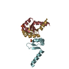
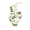
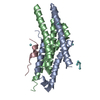
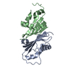
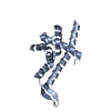
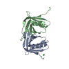
- PDB-5ott: Extracellular domain of GLP-1 receptor in complex with exendin-4 ... -
+ Open data

- Basic information
| Entry | Database: PDB / ID: 5ott | ||||||
|---|---|---|---|---|---|---|---|
| Title | Extracellular domain of GLP-1 receptor in complex with exendin-4 variant Gly2Hcs/Thr5Hcs | ||||||
 Components Components |
| ||||||
 Keywords Keywords | SIGNALING PROTEIN / glucagon-like peptide 1 / GPCR / cyclic peptides | ||||||
| Function / homology |  Function and homology information Function and homology informationglucagon-like peptide 1 receptor activity / glucagon receptor activity / positive regulation of blood pressure / hormone secretion / post-translational protein targeting to membrane, translocation / response to psychosocial stress / regulation of heart contraction / peptide hormone binding / activation of adenylate cyclase activity / negative regulation of blood pressure ...glucagon-like peptide 1 receptor activity / glucagon receptor activity / positive regulation of blood pressure / hormone secretion / post-translational protein targeting to membrane, translocation / response to psychosocial stress / regulation of heart contraction / peptide hormone binding / activation of adenylate cyclase activity / negative regulation of blood pressure / hormone activity / regulation of blood pressure / Glucagon-type ligand receptors / transmembrane signaling receptor activity / Glucagon-like Peptide-1 (GLP1) regulates insulin secretion / toxin activity / adenylate cyclase-activating G protein-coupled receptor signaling pathway / positive regulation of cytosolic calcium ion concentration / G alpha (s) signalling events / learning or memory / cell surface receptor signaling pathway / extracellular region / membrane / plasma membrane Similarity search - Function | ||||||
| Biological species |  Homo sapiens (human) Homo sapiens (human) Heloderma suspectum (Gila monster) Heloderma suspectum (Gila monster) | ||||||
| Method |  X-RAY DIFFRACTION / MOLECULAR REPLACEMENT / Resolution: 1.92 Å X-RAY DIFFRACTION / MOLECULAR REPLACEMENT / Resolution: 1.92 Å | ||||||
 Authors Authors | Mortensen, S. | ||||||
 Citation Citation | Journal: Biochemistry / Year: 2018 Title: alpha-Helix or beta-Turn? An Investigation into N-Terminally Constrained Analogues of Glucagon-like Peptide 1 (GLP-1) and Exendin-4. Authors: Oddo, A. / Mortensen, S. / Thogersen, H. / De Maria, L. / Hennen, S. / McGuire, J.N. / Kofoed, J. / Linderoth, L. / Reedtz-Runge, S. | ||||||
| History |
|
- Structure visualization
| Structure viewer | Molecule: MolmilJmol/JSmol |
|---|
- Downloads & links
-Download
| PDBx/mmCIF format | 5ott.cif.gz | 45.8 KB | Display | PDBx/mmCIF format |
|---|---|---|---|---|
| PDB format | pdb5ott.ent.gz | 30.5 KB | Display | PDB format |
| PDBx/mmJSON format | 5ott.json.gz | Tree view | PDBx/mmJSON format | |
| Others |  Other downloads Other downloads |
-Validation report
| Arichive directory | https://data.pdbj.org/pub/pdb/validation_reports/ot/5ottftp://data.pdbj.org/pub/pdb/validation_reports/ot/5ott | HTTPS FTP |
|---|
-Related structure data
| Related structure data |  5otuC  5otvC  5otwC  5otxC  4zgmS S: Starting model for refinement C: citing same article ( |
|---|---|
| Similar structure data |


-Links
 PDBj
PDBj


- Assembly
| Deposited unit | 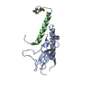
| |||||||||
|---|---|---|---|---|---|---|---|---|---|---|
| 1 |
| |||||||||
| Unit cell |
| |||||||||
| Components on special symmetry positions |
|
-Components
| #1: Protein | Mass: 13547.892 Da / Num. of mol.: 1 / Fragment: extracellular domain, UNP residues 24-139 Source method: isolated from a genetically manipulated source Source: (gene. exp.) Homo sapiens (human) / Gene: GLP1R / Production host:  |
|---|---|
| #2: Protein/peptide | Mass: 4267.772 Da / Num. of mol.: 1 / Mutation: G2HCS, T5HCS / Source method: obtained synthetically / Source: (synth.) Heloderma suspectum (Gila monster) / References: UniProt: P26349 |
| #3: Water | ChemComp-HOH /  Mass: 18.015 Da / Num. of mol.: 118 / Source method: isolated from a natural source / Formula: H2O Mass: 18.015 Da / Num. of mol.: 118 / Source method: isolated from a natural source / Formula: H2O |
| Has protein modification | Y |
-Experimental details
-Experiment
| Experiment | Method: X-RAY DIFFRACTION / Number of used crystals: 1 |
|---|
- Sample preparation
| Crystal | Density Matthews: 2.71 Å3/Da / Density % sol: 54.53 % |
|---|---|
| Crystal grow | Temperature: 293 K / Method: vapor diffusion, sitting drop Details: 0.1 M Imidazole pH 6.5, 1.0 M Sodium acetate trihydrate |
-Data collection
| Diffraction | Mean temperature: 100 K |
|---|---|
| Diffraction source | Source: ROTATING ANODE / Type: RIGAKU FR-X / Wavelength: 1.54187 Å |
| Detector | Type: DECTRIS PILATUS3 R 1M / Detector: PIXEL / Date: Feb 24, 2017 |
| Radiation | Protocol: SINGLE WAVELENGTH / Monochromatic (M) / Laue (L): M / Scattering type: x-ray |
| Radiation wavelength | Wavelength: 1.54187 Å / Relative weight: 1 |
| Reflection | Resolution: 1.92→47.382 Å / Num. obs: 15019 / % possible obs: 99 % / Redundancy: 12.9 % / CC1/2: 0.999 / Rmerge(I) obs: 0.103 / Rpim(I) all: 0.03 / Rsym value: 0.108 / Net I/σ(I): 19.8 |
| Reflection shell | Resolution: 1.92→1.95 Å / Redundancy: 12.66 % / Rmerge(I) obs: 1.145 / Mean I/σ(I) obs: 2.3 / Num. measured obs: 9519 / Num. unique all: 752 / Rpim(I) all: 0.33 / Rrim(I) all: 1.193 / % possible all: 97.41 |
- Processing
| Software |
| ||||||||||||||||||||||||||||||||||||||||||||||||||||||||||||||||||||||||||||||||||||||||||||||||||||||||||||||||||||||||||||||||||||||||||||
|---|---|---|---|---|---|---|---|---|---|---|---|---|---|---|---|---|---|---|---|---|---|---|---|---|---|---|---|---|---|---|---|---|---|---|---|---|---|---|---|---|---|---|---|---|---|---|---|---|---|---|---|---|---|---|---|---|---|---|---|---|---|---|---|---|---|---|---|---|---|---|---|---|---|---|---|---|---|---|---|---|---|---|---|---|---|---|---|---|---|---|---|---|---|---|---|---|---|---|---|---|---|---|---|---|---|---|---|---|---|---|---|---|---|---|---|---|---|---|---|---|---|---|---|---|---|---|---|---|---|---|---|---|---|---|---|---|---|---|---|---|---|
| Refinement | Method to determine structure: MOLECULAR REPLACEMENT Starting model: 4ZGM Resolution: 1.92→47.382 Å / SU ML: 0.17 / Cross valid method: FREE R-VALUE / σ(F): 1.34 / Phase error: 21.35
| ||||||||||||||||||||||||||||||||||||||||||||||||||||||||||||||||||||||||||||||||||||||||||||||||||||||||||||||||||||||||||||||||||||||||||||
| Solvent computation | Shrinkage radii: 0.9 Å / VDW probe radii: 1.11 Å | ||||||||||||||||||||||||||||||||||||||||||||||||||||||||||||||||||||||||||||||||||||||||||||||||||||||||||||||||||||||||||||||||||||||||||||
| Refinement step | Cycle: LAST / Resolution: 1.92→47.382 Å
| ||||||||||||||||||||||||||||||||||||||||||||||||||||||||||||||||||||||||||||||||||||||||||||||||||||||||||||||||||||||||||||||||||||||||||||
| Refine LS restraints |
| ||||||||||||||||||||||||||||||||||||||||||||||||||||||||||||||||||||||||||||||||||||||||||||||||||||||||||||||||||||||||||||||||||||||||||||
| LS refinement shell |
|
