 Movie
Movie Controller

[English] 日本語
 Yorodumi
- PDB-3hs2: Crystal structure of PHD truncated to residue 57 in an orthorhomb... -
+ Open data

- Basic information
| Entry | Database: PDB / ID: 3hs2 | ||||||
|---|---|---|---|---|---|---|---|
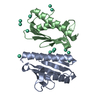
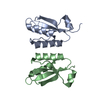
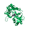
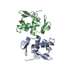
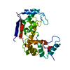
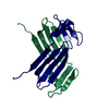
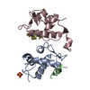
| Title | Crystal structure of PHD truncated to residue 57 in an orthorhombic space group | ||||||
 Components Components | Prevent host death protein | ||||||
 Keywords Keywords | ANTITOXIN / Prevent Host Death / PHD / intrinsic disorder / Doc / toxin-antitoxin | ||||||
| Function / homology |  Function and homology information Function and homology informationtoxin sequestering activity / DNA-binding transcription repressor activity / protein-DNA complex / sequence-specific DNA binding / negative regulation of DNA-templated transcription / protein homodimerization activity / DNA binding Similarity search - Function | ||||||
| Biological species |  Enterobacteria phage P1 (virus) Enterobacteria phage P1 (virus) | ||||||
| Method |  X-RAY DIFFRACTION / SYNCHROTRON / MOLECULAR REPLACEMENT / Resolution: 2.2 Å X-RAY DIFFRACTION / SYNCHROTRON / MOLECULAR REPLACEMENT / Resolution: 2.2 Å | ||||||
 Authors Authors | Garcia-Pino, A. / Loris, R. | ||||||
 Citation Citation | Journal: Cell(Cambridge,Mass.) / Year: 2010 Title: Allostery and intrinsic disorder mediate transcription regulation by conditional cooperativity. Authors: Garcia-Pino, A. / Balasubramanian, S. / Wyns, L. / Gazit, E. / De Greve, H. / Magnuson, R.D. / Charlier, D. / van Nuland, N.A. / Loris, R. | ||||||
| History |
|
- Structure visualization
| Structure viewer | Molecule: MolmilJmol/JSmol |
|---|
- Downloads & links
-Download
| PDBx/mmCIF format | 3hs2.cif.gz | 92.7 KB | Display | PDBx/mmCIF format |
|---|---|---|---|---|
| PDB format | pdb3hs2.ent.gz | 72.1 KB | Display | PDB format |
| PDBx/mmJSON format | 3hs2.json.gz | Tree view | PDBx/mmJSON format | |
| Others |  Other downloads Other downloads |
-Validation report
| Arichive directory | https://data.pdbj.org/pub/pdb/validation_reports/hs/3hs2ftp://data.pdbj.org/pub/pdb/validation_reports/hs/3hs2 | HTTPS FTP |
|---|
-Related structure data
| Related structure data |  3hrySC  3k33C S: Starting model for refinement C: citing same article ( |
|---|---|
| Similar structure data |


-Links
 PDBj
PDBj- Assembly
| Deposited unit | 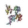
| ||||||||
|---|---|---|---|---|---|---|---|---|---|
| 1 | 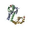
| ||||||||
| 2 | 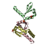
| ||||||||
| 3 | 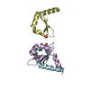
| ||||||||
| 4 | 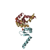
| ||||||||
| Unit cell |
|
-Components
| #1: Protein | Mass: 6394.143 Da / Num. of mol.: 8 / Fragment: N-terminal domain: UNP residues 1-58 Source method: isolated from a genetically manipulated source Source: (gene. exp.) Enterobacteria phage P1 (virus) / Gene: phd / Plasmid: pET21b / Production host:  #2: Chemical | ChemComp-SO4 /   Mass: 96.063 Da / Num. of mol.: 6 / Source method: obtained synthetically / Formula: SO4 Mass: 96.063 Da / Num. of mol.: 6 / Source method: obtained synthetically / Formula: SO4#3: Water | ChemComp-HOH / |  Mass: 18.015 Da / Num. of mol.: 83 / Source method: isolated from a natural source / Formula: H2O Mass: 18.015 Da / Num. of mol.: 83 / Source method: isolated from a natural source / Formula: H2O |
|---|
-Experimental details
-Experiment
| Experiment | Method: X-RAY DIFFRACTION / Number of used crystals: 1 |
|---|
- Sample preparation
| Crystal | Density Matthews: 1.96 Å3/Da / Density % sol: 37.25 % |
|---|---|
| Crystal grow | Temperature: 293 K / Method: vapor diffusion, hanging drop / pH: 6.5 Details: 18% PEG 8000, 0.2M LiSO4, 0.1M Sodium cacodylate, pH 6.5, VAPOR DIFFUSION, HANGING DROP, temperature 293K |
-Data collection
| Diffraction | Mean temperature: 100 K |
|---|---|
| Diffraction source | Source: SYNCHROTRON / Site: SOLEIL  / Beamline: PROXIMA 1 / Wavelength: 0.9801 Å / Beamline: PROXIMA 1 / Wavelength: 0.9801 Å |
| Detector | Type: ADSC QUANTUM 315r / Detector: CCD / Date: Feb 14, 2009 / Details: mirrors |
| Radiation | Protocol: SINGLE WAVELENGTH / Monochromatic (M) / Laue (L): M / Scattering type: x-ray |
| Radiation wavelength | Wavelength: 0.9801 Å / Relative weight: 1 |
| Reflection | Resolution: 2.2→34.25 Å / Num. all: 20810 / Num. obs: 20810 / % possible obs: 99.1 % / Observed criterion σ(F): 0 / Observed criterion σ(I): 0 / Redundancy: 8 % / Biso Wilson estimate: 36.4 Å2 / Rmerge(I) obs: 0.11 / Rsym value: 0.11 / Net I/σ(I): 9 |
| Reflection shell | Resolution: 2.2→2.28 Å / Redundancy: 4.8 % / Rmerge(I) obs: 0.321 / Mean I/σ(I) obs: 6.87 / Num. unique all: 2025 / Rsym value: 0.321 / % possible all: 99.9 |
- Processing
| Software |
| ||||||||||||||||||||||||||||||||||||||||||||||||||||||||||||||||||
|---|---|---|---|---|---|---|---|---|---|---|---|---|---|---|---|---|---|---|---|---|---|---|---|---|---|---|---|---|---|---|---|---|---|---|---|---|---|---|---|---|---|---|---|---|---|---|---|---|---|---|---|---|---|---|---|---|---|---|---|---|---|---|---|---|---|---|---|
| Refinement | Method to determine structure: MOLECULAR REPLACEMENT Starting model: PDB entry 3HRY Resolution: 2.2→34.25 Å / SU ML: 1.74 / σ(F): 0 / σ(I): 0 / Stereochemistry target values: ML
| ||||||||||||||||||||||||||||||||||||||||||||||||||||||||||||||||||
| Solvent computation | Shrinkage radii: 0.9 Å / VDW probe radii: 1.11 Å / Solvent model: FLAT BULK SOLVENT MODEL / Bsol: 56.099 Å2 / ksol: 0.379 e/Å3 | ||||||||||||||||||||||||||||||||||||||||||||||||||||||||||||||||||
| Refinement step | Cycle: LAST / Resolution: 2.2→34.25 Å
| ||||||||||||||||||||||||||||||||||||||||||||||||||||||||||||||||||
| Refine LS restraints |
| ||||||||||||||||||||||||||||||||||||||||||||||||||||||||||||||||||
| LS refinement shell | Refine-ID: X-RAY DIFFRACTION / Total num. of bins used: 10
|
