 Movie
Movie Controller


+ Open data

- Basic information
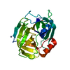
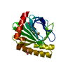
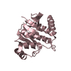
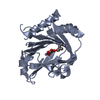
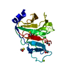
| Entry | Database: PDB / ID: 5onj | ||||||
|---|---|---|---|---|---|---|---|
| Title | YnDL in Complex with 5 amino acid (PGA) complex | ||||||
 Components Components |
| ||||||
 Keywords Keywords | HYDROLASE / PGA-HYDROLASE | ||||||
| Function / homology | Poly-gamma-glutamate hydrolase / Poly-gamma-glutamate hydrolase, zinc-binding motif superfamily / Poly-gamma-glutamate hydrolase / hydrolase activity / plasma membrane / Gamma-polyglutamate hydrolase PghL / UPF0714 protein YndL Function and homology information Function and homology information | ||||||
| Biological species |  synthetic construct (others) | ||||||
| Method |  X-RAY DIFFRACTION / SYNCHROTRON / MOLECULAR REPLACEMENT / Resolution: 1.7 Å X-RAY DIFFRACTION / SYNCHROTRON / MOLECULAR REPLACEMENT / Resolution: 1.7 Å | ||||||
 Authors Authors | Ramaswamy, S. / Rasheed, M. / Morelli, C. / Calvio, C. / Sutton, B. / Pastore, A. | ||||||
 Citation Citation | Journal: Febs J. / Year: 2018 Title: The structure of PghL hydrolase bound to its substrate poly-gamma-glutamate. Authors: Ramaswamy, S. / Rasheed, M. / Morelli, C.F. / Calvio, C. / Sutton, B.J. / Pastore, A. #1: Journal: To Be PublishedTitle: Poly-gamma-glutamate hydrolases structure: a potential strategy against Staphylococcus infectivity Authors: Ramaswamy, S. / Rasheed, M. / Morelli, C. / Calvio, C. / Sutton, B. / Pastore, A. | ||||||
| History |
|
- Structure visualization
| Structure viewer | Molecule: MolmilJmol/JSmol |
|---|
- Downloads & links
-Download
| PDBx/mmCIF format | 5onj.cif.gz | 62.8 KB | Display | PDBx/mmCIF format |
|---|---|---|---|---|
| PDB format | pdb5onj.ent.gz | 44.3 KB | Display | PDB format |
| PDBx/mmJSON format | 5onj.json.gz | Tree view | PDBx/mmJSON format | |
| Others |  Other downloads Other downloads |
-Validation report
| Arichive directory | https://data.pdbj.org/pub/pdb/validation_reports/on/5onjftp://data.pdbj.org/pub/pdb/validation_reports/on/5onj | HTTPS FTP |
|---|
-Related structure data
| Related structure data | 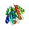 5onkC 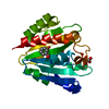 5onlC  6hriC 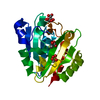 6hrjC 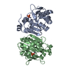 3a9lS S: Starting model for refinement C: citing same article ( |
|---|---|
| Similar structure data |


-Links
 PDBj
PDBj- Assembly
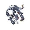
| Deposited unit | 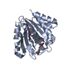
| ||||||||
|---|---|---|---|---|---|---|---|---|---|
| 1 |
| ||||||||
| Unit cell |
|
-Components
| #1: Protein | Mass: 23985.703 Da / Num. of mol.: 1 Source method: isolated from a genetically manipulated source Source: (gene. exp.) |
|---|---|
| #2: Protein/peptide | ( Mass: 792.698 Da / Num. of mol.: 1 / Source method: obtained synthetically / Source: (synth.) synthetic construct (others) |
| #3: Chemical | ChemComp-ZN /   Mass: 65.409 Da / Num. of mol.: 1 / Source method: obtained synthetically / Formula: Zn Mass: 65.409 Da / Num. of mol.: 1 / Source method: obtained synthetically / Formula: Zn |
| #4: Water | ChemComp-HOH /  Mass: 18.015 Da / Num. of mol.: 228 / Source method: isolated from a natural source / Formula: H2O Mass: 18.015 Da / Num. of mol.: 228 / Source method: isolated from a natural source / Formula: H2O |
| Has protein modification | Y |
-Experimental details
-Experiment
| Experiment | Method: X-RAY DIFFRACTION / Number of used crystals: 1 |
|---|
- Sample preparation
| Crystal | Density Matthews: 1.86 Å3/Da / Density % sol: 33.94 % |
|---|---|
| Crystal grow | Temperature: 291 K / Method: vapor diffusion, sitting drop / Details: 1M Sodium Acetate, 5% gamma-PGA, 20% PEG 3350 |
-Data collection
| Diffraction | Mean temperature: 100 K |
|---|---|
| Diffraction source | Source: SYNCHROTRON / Site: Diamond  / Beamline: I03 / Wavelength: 1 Å / Beamline: I03 / Wavelength: 1 Å |
| Detector | Type: DECTRIS PILATUS3 6M / Detector: PIXEL / Date: Jul 10, 2017 |
| Radiation | Protocol: SINGLE WAVELENGTH / Monochromatic (M) / Laue (L): M / Scattering type: x-ray |
| Radiation wavelength | Wavelength: 1 Å / Relative weight: 1 |
| Reflection | Resolution: 1.7→15.75 Å / Num. obs: 20613 / % possible obs: 99 % / Redundancy: 1.9 % / Biso Wilson estimate: 14.34 Å2 / Net I/σ(I): 24.58 |
- Processing
| Software |
| |||||||||||||||||||||||||||||||||||||||||||||||||||||||||||||||
|---|---|---|---|---|---|---|---|---|---|---|---|---|---|---|---|---|---|---|---|---|---|---|---|---|---|---|---|---|---|---|---|---|---|---|---|---|---|---|---|---|---|---|---|---|---|---|---|---|---|---|---|---|---|---|---|---|---|---|---|---|---|---|---|---|
| Refinement | Method to determine structure: MOLECULAR REPLACEMENT Starting model: 3A9L Resolution: 1.7→15.75 Å / SU ML: 0.17 / Cross valid method: THROUGHOUT / σ(F): 1.34 / Phase error: 20.28
| |||||||||||||||||||||||||||||||||||||||||||||||||||||||||||||||
| Solvent computation | Shrinkage radii: 0.9 Å / VDW probe radii: 1.11 Å | |||||||||||||||||||||||||||||||||||||||||||||||||||||||||||||||
| Refinement step | Cycle: LAST / Resolution: 1.7→15.75 Å
| |||||||||||||||||||||||||||||||||||||||||||||||||||||||||||||||
| Refine LS restraints |
| |||||||||||||||||||||||||||||||||||||||||||||||||||||||||||||||
| LS refinement shell |
|
