 Movie
Movie Controller

[English] 日本語
 Yorodumi
- PDB-4qkd: Crystal structure of human ALKBH7 in complex with alpha-ketogluta... -
+ Open data

- Basic information
| Entry | Database: PDB / ID: 4qkd | ||||||
|---|---|---|---|---|---|---|---|
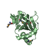
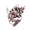
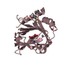
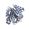
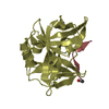
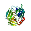
| Title | Crystal structure of human ALKBH7 in complex with alpha-ketoglutarate and Mn(II) | ||||||
 Components Components | Alpha-ketoglutarate-dependent dioxygenase alkB homolog 7, mitochondrial | ||||||
 Keywords Keywords | OXIDOREDUCTASE / alkbh7 / DIOXYGENASE / METAL-BINDING / programmed necrosis / fat metabolism | ||||||
| Function / homology |  Function and homology information Function and homology informationtRNA demethylase activity / oxidative RNA demethylation / regulation of lipid storage / oxidative RNA demethylase activity / regulation of mitochondrial membrane permeability involved in programmed necrotic cell death / Oxidoreductases; Acting on paired donors, with incorporation or reduction of molecular oxygen; With 2-oxoglutarate as one donor, and incorporation of one atom of oxygen into each donor / fatty acid metabolic process / mitochondrial matrix / DNA damage response / mitochondrion / metal ion binding Similarity search - Function | ||||||
| Biological species |  Homo sapiens (human) Homo sapiens (human) | ||||||
| Method |  X-RAY DIFFRACTION / SYNCHROTRON / MOLECULAR REPLACEMENT / Resolution: 1.35 Å X-RAY DIFFRACTION / SYNCHROTRON / MOLECULAR REPLACEMENT / Resolution: 1.35 Å | ||||||
 Authors Authors | Wang, G. / Chen, Z. | ||||||
 Citation Citation | Journal: J.Biol.Chem. / Year: 2014 Title: The atomic resolution structure of human AlkB homolog 7 (ALKBH7), a key protein for programmed necrosis and fat metabolism Authors: Wang, G. / He, Q. / Feng, C. / Liu, Y. / Deng, Z. / Qi, X. / Wu, W. / Mei, P. / Chen, Z. | ||||||
| History |
|
- Structure visualization
| Structure viewer | Molecule: MolmilJmol/JSmol |
|---|
- Downloads & links
-Download
| PDBx/mmCIF format | 4qkd.cif.gz | 267 KB | Display | PDBx/mmCIF format |
|---|---|---|---|---|
| PDB format | pdb4qkd.ent.gz | 214.3 KB | Display | PDB format |
| PDBx/mmJSON format | 4qkd.json.gz | Tree view | PDBx/mmJSON format | |
| Others |  Other downloads Other downloads |
-Validation report
| Arichive directory | https://data.pdbj.org/pub/pdb/validation_reports/qk/4qkdftp://data.pdbj.org/pub/pdb/validation_reports/qk/4qkd | HTTPS FTP |
|---|
-Related structure data
| Related structure data |  4qkbSC 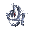 4qkfC C: citing same article ( S: Starting model for refinement |
|---|---|
| Similar structure data |


-Links
 PDBj
PDBj

- Assembly
| Deposited unit | 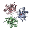
| ||||||||
|---|---|---|---|---|---|---|---|---|---|
| 1 | 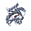
| ||||||||
| 2 | 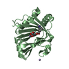
| ||||||||
| 3 | 
| ||||||||
| 4 |
| ||||||||
| Unit cell |
|
-Components
| #1: Protein | Mass: 22591.590 Da / Num. of mol.: 3 / Fragment: UNP RESIDUES 17-215 / Mutation: Q90R Source method: isolated from a genetically manipulated source Source: (gene. exp.) Homo sapiens (human) / Gene: ABH7, ALKBH7, SPATA11, UNQ6002/PRO34564 / Production host:  References: UniProt: Q9BT30, Oxidoreductases; Acting on paired donors, with incorporation or reduction of molecular oxygen; With 2-oxoglutarate as one donor, and incorporation of one atom of oxygen into each donor #2: Chemical | ChemComp-MN /   Mass: 54.938 Da / Num. of mol.: 7 / Source method: obtained synthetically / Formula: Mn Mass: 54.938 Da / Num. of mol.: 7 / Source method: obtained synthetically / Formula: Mn#3: Chemical |   Mass: 146.098 Da / Num. of mol.: 3 / Source method: obtained synthetically / Formula: C5H6O5 Mass: 146.098 Da / Num. of mol.: 3 / Source method: obtained synthetically / Formula: C5H6O5#4: Water | ChemComp-HOH / |  Mass: 18.015 Da / Num. of mol.: 731 / Source method: isolated from a natural source / Formula: H2O Mass: 18.015 Da / Num. of mol.: 731 / Source method: isolated from a natural source / Formula: H2OHas protein modification | Y | |
|---|
-Experimental details
-Experiment
| Experiment | Method: X-RAY DIFFRACTION / Number of used crystals: 3 |
|---|
- Sample preparation
| Crystal | Density Matthews: 2.31 Å3/Da / Density % sol: 46.8 % |
|---|---|
| Crystal grow | Temperature: 277 K / Method: evaporation / pH: 6 / Details: PEG pH6, EVAPORATION, temperature 277K |
-Data collection
| Diffraction | Mean temperature: 130 K | ||||||||||||||||||||
|---|---|---|---|---|---|---|---|---|---|---|---|---|---|---|---|---|---|---|---|---|---|
| Diffraction source | Source: SYNCHROTRON / Site: SSRF  / Beamline: BL17U / Wavelength: 0.9793 Å / Beamline: BL17U / Wavelength: 0.9793 Å | ||||||||||||||||||||
| Detector | Type: ADSC QUANTUM 315 / Detector: CCD / Date: Oct 13, 2012 | ||||||||||||||||||||
| Radiation | Protocol: SINGLE WAVELENGTH / Monochromatic (M) / Laue (L): M / Scattering type: x-ray | ||||||||||||||||||||
| Radiation wavelength | Wavelength: 0.9793 Å / Relative weight: 1 | ||||||||||||||||||||
| Reflection twin |
| ||||||||||||||||||||
| Reflection | Resolution: 1.35→50 Å / Num. all: 135021 / Num. obs: 124116 / % possible obs: 91.9 % / Observed criterion σ(F): 3 / Observed criterion σ(I): 3 | ||||||||||||||||||||
| Reflection shell | Resolution: 1.35→1.37 Å / % possible all: 61 |
- Processing
| Software |
| ||||||||||||||||||||||||||||||||||||||||||||||||||||||||||||||||||||||||||||||||||||||||||||||||||||||||||||||||||||||||
|---|---|---|---|---|---|---|---|---|---|---|---|---|---|---|---|---|---|---|---|---|---|---|---|---|---|---|---|---|---|---|---|---|---|---|---|---|---|---|---|---|---|---|---|---|---|---|---|---|---|---|---|---|---|---|---|---|---|---|---|---|---|---|---|---|---|---|---|---|---|---|---|---|---|---|---|---|---|---|---|---|---|---|---|---|---|---|---|---|---|---|---|---|---|---|---|---|---|---|---|---|---|---|---|---|---|---|---|---|---|---|---|---|---|---|---|---|---|---|---|---|---|
| Refinement | Method to determine structure: MOLECULAR REPLACEMENT Starting model: 4QKB Resolution: 1.35→33.4 Å / Cor.coef. Fo:Fc: 0.968 / Cor.coef. Fo:Fc free: 0.964 / SU B: 1.788 / SU ML: 0.035 / Cross valid method: THROUGHOUT / σ(F): 3 / ESU R: 0.012 / ESU R Free: 0.01 / Stereochemistry target values: MAXIMUM LIKELIHOOD / Details: HYDROGENS HAVE BEEN ADDED IN THE RIDING POSITIONS
| ||||||||||||||||||||||||||||||||||||||||||||||||||||||||||||||||||||||||||||||||||||||||||||||||||||||||||||||||||||||||
| Solvent computation | Ion probe radii: 0.8 Å / Shrinkage radii: 0.8 Å / VDW probe radii: 1.2 Å / Solvent model: MASK | ||||||||||||||||||||||||||||||||||||||||||||||||||||||||||||||||||||||||||||||||||||||||||||||||||||||||||||||||||||||||
| Displacement parameters | Biso mean: 17.989 Å2
| ||||||||||||||||||||||||||||||||||||||||||||||||||||||||||||||||||||||||||||||||||||||||||||||||||||||||||||||||||||||||
| Refinement step | Cycle: LAST / Resolution: 1.35→33.4 Å
| ||||||||||||||||||||||||||||||||||||||||||||||||||||||||||||||||||||||||||||||||||||||||||||||||||||||||||||||||||||||||
| Refine LS restraints |
| ||||||||||||||||||||||||||||||||||||||||||||||||||||||||||||||||||||||||||||||||||||||||||||||||||||||||||||||||||||||||
| LS refinement shell | Resolution: 1.35→1.385 Å / Total num. of bins used: 20
|
