tRNA demethylase activity / oxidative RNA demethylation / regulation of lipid storage / oxidative RNA demethylase activity / regulation of mitochondrial membrane permeability involved in programmed necrotic cell death / Oxidoreductases; Acting on paired donors, with incorporation or reduction of molecular oxygen; With 2-oxoglutarate as one donor, and incorporation of one atom of oxygen into each donor / fatty acid metabolic process / transferase activity / mitochondrial matrix / DNA damage response ...tRNA demethylase activity / oxidative RNA demethylation / regulation of lipid storage / oxidative RNA demethylase activity / regulation of mitochondrial membrane permeability involved in programmed necrotic cell death / Oxidoreductases; Acting on paired donors, with incorporation or reduction of molecular oxygen; With 2-oxoglutarate as one donor, and incorporation of one atom of oxygen into each donor / fatty acid metabolic process / transferase activity / mitochondrial matrix / DNA damage response / mitochondrion / metal ion binding Similarity search - Function
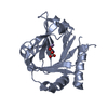
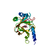
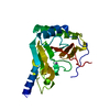
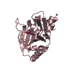
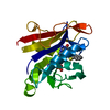
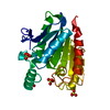
Alpha-ketoglutarate-dependentdioxygenasealkBhomolog7, mitochondrial / Alkylated DNA repair protein alkB homolog 7 / Spermatogenesis cell proliferation-related protein / ...Alkylated DNA repair protein alkB homolog 7 / Spermatogenesis cell proliferation-related protein / Spermatogenesis-associated protein 11
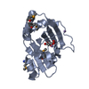
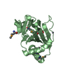
Mass: 22804.123 Da / Num. of mol.: 3 / Fragment: UNP RESIDUES 17-215 / Mutation: Q90R, R86M and L89M Source method: isolated from a genetically manipulated source Source: (gene. exp.) Homo sapiens (human) / Gene: ABH7, ALKBH7, SPATA11, UNQ6002/PRO34564 / Production host: Escherichia coli (E. coli) References: UniProt: Q9BT30, Oxidoreductases; Acting on paired donors, with incorporation or reduction of molecular oxygen; With 2-oxoglutarate as one donor, and incorporation of one atom of oxygen into each donor
Method to determine structure: MAD / Resolution: 2.6→50 Å / Cor.coef. Fo:Fc: 0.927 / Cor.coef. Fo:Fc free: 0.919 / SU B: 13.736 / SU ML: 0.282 / Cross valid method: THROUGHOUT / σ(F): 3 / ESU R Free: 0.055 / Stereochemistry target values: MAXIMUM LIKELIHOOD / Details: HYDROGENS HAVE BEEN USED IF PRESENT IN THE INPUT
Rfactor
Num. reflection
% reflection
Selection details
Rfree
0.18103
909
5.2 %
RANDOM
Rwork
0.15769
-
-
-
all
0.1589
17632
-
-
obs
0.1589
16706
90.07 %
-
Solvent computation
Ion probe radii: 0.8 Å / Shrinkage radii: 0.8 Å / VDW probe radii: 1.2 Å / Solvent model: MASK
Displacement parameters
Biso mean: 27.651 Å2
Baniso -1
Baniso -2
Baniso -3
1-
-29.06 Å2
-0 Å2
-0.78 Å2
2-
-
44.21 Å2
0 Å2
3-
-
-
-15.14 Å2
Refinement step
Cycle: LAST / Resolution: 2.6→50 Å
Protein
Nucleic acid
Ligand
Solvent
Total
Num. atoms
4476
0
37
54
4567
Refine LS restraints
Refine-ID
Type
Dev ideal
Dev ideal target
Number
X-RAY DIFFRACTION
r_bond_refined_d
0.007
0.019
4605
X-RAY DIFFRACTION
r_angle_refined_deg
1.152
1.981
6221
X-RAY DIFFRACTION
r_dihedral_angle_1_deg
6.024
5
564
X-RAY DIFFRACTION
r_dihedral_angle_2_deg
31.104
21.232
203
X-RAY DIFFRACTION
r_dihedral_angle_3_deg
14.684
15
732
X-RAY DIFFRACTION
r_dihedral_angle_4_deg
17.024
15
59
X-RAY DIFFRACTION
r_chiral_restr
0.07
0.2
675
X-RAY DIFFRACTION
r_gen_planes_refined
0.004
0.021
3499
LS refinement shell
Resolution: 2.596→2.663 Å / Total num. of bins used: 20
Rfactor
Num. reflection
% reflection
Rfree
0.243
39
-
Rwork
0.257
667
-
obs
-
-
48.86 %
+
About Yorodumi
-
News
-
Feb 9, 2022. New format data for meta-information of EMDB entries
New format data for meta-information of EMDB entries
Version 3 of the EMDB header file is now the official format.
The previous official version 1.9 will be removed from the archive.
In the structure databanks used in Yorodumi, some data are registered as the other names, "COVID-19 virus" and "2019-nCoV". Here are the details of the virus and the list of structure data.
Jan 31, 2019. EMDB accession codes are about to change! (news from PDBe EMDB page)
EMDB accession codes are about to change! (news from PDBe EMDB page)
The allocation of 4 digits for EMDB accession codes will soon come to an end. Whilst these codes will remain in use, new EMDB accession codes will include an additional digit and will expand incrementally as the available range of codes is exhausted. The current 4-digit format prefixed with “EMD-” (i.e. EMD-XXXX) will advance to a 5-digit format (i.e. EMD-XXXXX), and so on. It is currently estimated that the 4-digit codes will be depleted around Spring 2019, at which point the 5-digit format will come into force.
The EM Navigator/Yorodumi systems omit the EMD- prefix.
Related info.:Q: What is EMD? / ID/Accession-code notation in Yorodumi/EM Navigator
Yorodumi is a browser for structure data from EMDB, PDB, SASBDB, etc.
This page is also the successor to EM Navigator detail page, and also detail information page/front-end page for Omokage search.
The word "yorodu" (or yorozu) is an old Japanese word meaning "ten thousand". "mi" (miru) is to see.
Related info.:EMDB / PDB / SASBDB / Comparison of 3 databanks / Yorodumi Search / Aug 31, 2016. New EM Navigator & Yorodumi / Yorodumi Papers / Jmol/JSmol / Function and homology information / Changes in new EM Navigator and Yorodumi
 Movie
Movie Controller

 Yorodumi
 Open data
 Basic information
 Components
 Keywords
 Function and homology information
 Homo sapiens (human)
 X-RAY DIFFRACTION /
  Authors
 Citation
 Structure visualization
 Downloads & links
 Other downloads


 PDBj
PDBj

 Assembly
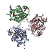
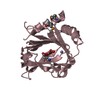


 Mass: 54.938 Da / Num. of mol.: 7 / Source method: obtained synthetically / Formula: Mn
 Mass: 146.098 Da / Num. of mol.: 3 / Source method: obtained synthetically / Formula: C5H6O5
 Mass: 18.015 Da / Num. of mol.: 54 / Source method: isolated from a natural source / Formula: H2O
 Sample preparation
 / Beamline: BL-5A / Wavelength: 0.97929, 0.97943
 Processing