 Movie
Movie Controller

[English] 日本語
 Yorodumi
- PDB-5omy: HIGH-SALT STRUCTURE OF PROTEIN KINASE CK2 CATALYTIC SUBUNIT (ISOF... -
+ Open data

- Basic information
| Entry | Database: PDB / ID: 5omy | ||||||
|---|---|---|---|---|---|---|---|
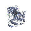
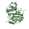
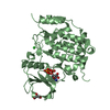
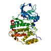
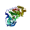
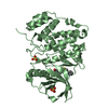
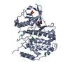
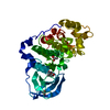
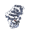
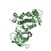
| Title | HIGH-SALT STRUCTURE OF PROTEIN KINASE CK2 CATALYTIC SUBUNIT (ISOFORM CK2ALPHA) IN COMPLEX WITH THE INDENOINDOLE-TYPE INHIBITOR 4P | ||||||
 Components Components | Casein kinase II subunit alpha | ||||||
 Keywords Keywords | TRANSFERASE / protein kinase CK2 / casein kinase 2 / indenoindole-type inhibitors | ||||||
| Function / homology |  Function and homology information Function and homology informationPhosphorylation and nuclear translocation of BMAL1 (ARNTL) and CLOCK / positive regulation of aggrephagy / regulation of chromosome separation / WNT mediated activation of DVL / Condensation of Prometaphase Chromosomes / protein kinase CK2 complex / symbiont-mediated disruption of host cell PML body / Phosphorylation and nuclear translocation of the CRY:PER:kinase complex / Regulation of CDH1 posttranslational processing and trafficking to plasma membrane / Receptor Mediated Mitophagy ...Phosphorylation and nuclear translocation of BMAL1 (ARNTL) and CLOCK / positive regulation of aggrephagy / regulation of chromosome separation / WNT mediated activation of DVL / Condensation of Prometaphase Chromosomes / protein kinase CK2 complex / symbiont-mediated disruption of host cell PML body / Phosphorylation and nuclear translocation of the CRY:PER:kinase complex / Regulation of CDH1 posttranslational processing and trafficking to plasma membrane / Receptor Mediated Mitophagy / Synthesis of PC / Sin3-type complex / negative regulation of signal transduction by p53 class mediator / RUNX1 interacts with co-factors whose precise effect on RUNX1 targets is not known / Maturation of hRSV A proteins / negative regulation of apoptotic signaling pathway / negative regulation of double-strand break repair via homologous recombination / positive regulation of Wnt signaling pathway / negative regulation of proteasomal ubiquitin-dependent protein catabolic process / Signal transduction by L1 / Hsp90 protein binding / PML body / Wnt signaling pathway / Regulation of PTEN stability and activity / kinase activity / positive regulation of protein catabolic process / KEAP1-NFE2L2 pathway / rhythmic process / double-strand break repair / Cooperation of PDCL (PhLP1) and TRiC/CCT in G-protein beta folding / positive regulation of cell growth / protein folding / Regulation of TP53 Activity through Phosphorylation / non-specific serine/threonine protein kinase / regulation of cell cycle / negative regulation of translation / protein stabilization / protein serine kinase activity / protein serine/threonine kinase activity / apoptotic process / positive regulation of cell population proliferation / DNA damage response / signal transduction / nucleoplasm / ATP binding / identical protein binding / nucleus / plasma membrane / cytosol Similarity search - Function | ||||||
| Biological species |  Homo sapiens (human) Homo sapiens (human) | ||||||
| Method |  X-RAY DIFFRACTION / SYNCHROTRON / MOLECULAR REPLACEMENT / Resolution: 1.95 Å X-RAY DIFFRACTION / SYNCHROTRON / MOLECULAR REPLACEMENT / Resolution: 1.95 Å | ||||||
 Authors Authors | Hochscherf, J. / Lindenblatt, D. / Witulski, B. / Birus, R. / Aichele, D. / Marminon, C. / Bouaziz, Z. / Le Borgne, M. / Jose, J. / Niefind, K. | ||||||
| Funding support |  Germany, 1items Germany, 1items
| ||||||
 Citation Citation | Journal: Pharmaceuticals (Basel) / Year: 2017 Title: Unexpected Binding Mode of a Potent Indeno[1,2-b]indole-Type Inhibitor of Protein Kinase CK2 Revealed by Complex Structures with the Catalytic Subunit CK2 alpha and Its Paralog CK2 alpha '. Authors: Hochscherf, J. / Lindenblatt, D. / Witulski, B. / Birus, R. / Aichele, D. / Marminon, C. / Bouaziz, Z. / Le Borgne, M. / Jose, J. / Niefind, K. | ||||||
| History |
|
- Structure visualization
| Structure viewer | Molecule: MolmilJmol/JSmol |
|---|
- Downloads & links
-Download
| PDBx/mmCIF format | 5omy.cif.gz | 160.2 KB | Display | PDBx/mmCIF format |
|---|---|---|---|---|
| PDB format | pdb5omy.ent.gz | 125.7 KB | Display | PDB format |
| PDBx/mmJSON format | 5omy.json.gz | Tree view | PDBx/mmJSON format | |
| Others |  Other downloads Other downloads |
-Validation report
| Arichive directory | https://data.pdbj.org/pub/pdb/validation_reports/om/5omyftp://data.pdbj.org/pub/pdb/validation_reports/om/5omy | HTTPS FTP |
|---|
-Related structure data
| Related structure data |  5oniC  5ooiC  2pvrS S: Starting model for refinement C: citing same article ( |
|---|---|
| Similar structure data |

-Links
 PDBj
PDBj


- Assembly
| Deposited unit | 
| ||||||||
|---|---|---|---|---|---|---|---|---|---|
| 1 |
| ||||||||
| Unit cell |
|
-Components
| #1: Protein | Mass: 45208.559 Da / Num. of mol.: 1 Source method: isolated from a genetically manipulated source Source: (gene. exp.) Homo sapiens (human) / Gene: CSNK2A1, CK2A1 / Production host:  References: UniProt: P68400, non-specific serine/threonine protein kinase | ||
|---|---|---|---|
| #2: Chemical | ChemComp-9YE /   Mass: 363.450 Da / Num. of mol.: 1 / Source method: obtained synthetically / Formula: C23H25NO3 / Feature type: SUBJECT OF INVESTIGATION Mass: 363.450 Da / Num. of mol.: 1 / Source method: obtained synthetically / Formula: C23H25NO3 / Feature type: SUBJECT OF INVESTIGATION | ||
| #3: Chemical | ChemComp-CL /   Mass: 35.453 Da / Num. of mol.: 5 / Source method: obtained synthetically / Formula: Cl Mass: 35.453 Da / Num. of mol.: 5 / Source method: obtained synthetically / Formula: Cl#4: Water | ChemComp-HOH / |  Mass: 18.015 Da / Num. of mol.: 122 / Source method: isolated from a natural source / Formula: H2O Mass: 18.015 Da / Num. of mol.: 122 / Source method: isolated from a natural source / Formula: H2O |
-Experimental details
-Experiment
| Experiment | Method: X-RAY DIFFRACTION / Number of used crystals: 1 |
|---|
- Sample preparation
| Crystal | Density Matthews: 1.94 Å3/Da / Density % sol: 36.66 % |
|---|---|
| Crystal grow | Temperature: 293.15 K / Method: vapor diffusion, sitting drop Details: 90 MIKROLITER ENZYME STOCK SOLUTION (6 MG/ML IN 500 MM NACL, 25 MM TRIS/HCL, PH 8.5) WAS MIXED WITH 10 MIKROLITER 4P STOCK SOLUTION (10 MM 4P IN DMSO). THIS MIXTURE WAS INCUBATED FOR 30 MIN ...Details: 90 MIKROLITER ENZYME STOCK SOLUTION (6 MG/ML IN 500 MM NACL, 25 MM TRIS/HCL, PH 8.5) WAS MIXED WITH 10 MIKROLITER 4P STOCK SOLUTION (10 MM 4P IN DMSO). THIS MIXTURE WAS INCUBATED FOR 30 MIN AT ROOM TEMPERATURE. THE RESERVOIR SOLUTION OF THE CRYSTALLIZATION EXPERIMENT WAS 4.2 M NACL, 0.1 M CITRIC ACID, PH 5.5. PRIOR TO EQUILIBRATION THE CRYSTALLIZATION DROP WAS COMPOSED OF 1 MIKROLITER RESERVOIR SOLUTION PLUS 1 MIKROLITER ENZYME/4P MIXTURE., VAPOR DIFFUSION, SITTING DROP, TEMPERATURE 293.15K |
-Data collection
| Diffraction | Mean temperature: 100 K |
|---|---|
| Diffraction source | Source: SYNCHROTRON / Site: SLS  / Beamline: X06DA / Wavelength: 0.97625 Å / Beamline: X06DA / Wavelength: 0.97625 Å |
| Detector | Type: DECTRIS PILATUS 2M-F / Detector: PIXEL / Date: Feb 3, 2017 |
| Radiation | Protocol: SINGLE WAVELENGTH / Monochromatic (M) / Laue (L): M / Scattering type: x-ray |
| Radiation wavelength | Wavelength: 0.97625 Å / Relative weight: 1 |
| Reflection | Resolution: 1.95→63.78 Å / Num. obs: 26747 / % possible obs: 99.94 % / Redundancy: 24.9 % / Biso Wilson estimate: 43.54 Å2 / Rmerge(I) obs: 0.09151 / Net I/σ(I): 20.77 |
| Reflection shell | Resolution: 1.95→2.02 Å / Redundancy: 26.1 % / Rmerge(I) obs: 2.281 / Mean I/σ(I) obs: 1.52 / Num. unique obs: 2639 / CC1/2: 0.64 / % possible all: 100 |
- Processing
| Software |
| |||||||||||||||||||||||||||||||||||||||||||||||||||||||||||||||||||||||||||||||||||||||||||||||||||||||||||||||||||||||||||||
|---|---|---|---|---|---|---|---|---|---|---|---|---|---|---|---|---|---|---|---|---|---|---|---|---|---|---|---|---|---|---|---|---|---|---|---|---|---|---|---|---|---|---|---|---|---|---|---|---|---|---|---|---|---|---|---|---|---|---|---|---|---|---|---|---|---|---|---|---|---|---|---|---|---|---|---|---|---|---|---|---|---|---|---|---|---|---|---|---|---|---|---|---|---|---|---|---|---|---|---|---|---|---|---|---|---|---|---|---|---|---|---|---|---|---|---|---|---|---|---|---|---|---|---|---|---|---|
| Refinement | Method to determine structure: MOLECULAR REPLACEMENT Starting model: 2PVR Resolution: 1.95→63.778 Å / SU ML: 0.25 / Cross valid method: FREE R-VALUE / σ(F): 1.33 / Phase error: 22.93
| |||||||||||||||||||||||||||||||||||||||||||||||||||||||||||||||||||||||||||||||||||||||||||||||||||||||||||||||||||||||||||||
| Solvent computation | Shrinkage radii: 0.9 Å / VDW probe radii: 1.11 Å | |||||||||||||||||||||||||||||||||||||||||||||||||||||||||||||||||||||||||||||||||||||||||||||||||||||||||||||||||||||||||||||
| Refinement step | Cycle: LAST / Resolution: 1.95→63.778 Å
| |||||||||||||||||||||||||||||||||||||||||||||||||||||||||||||||||||||||||||||||||||||||||||||||||||||||||||||||||||||||||||||
| Refine LS restraints |
| |||||||||||||||||||||||||||||||||||||||||||||||||||||||||||||||||||||||||||||||||||||||||||||||||||||||||||||||||||||||||||||
| LS refinement shell |
| |||||||||||||||||||||||||||||||||||||||||||||||||||||||||||||||||||||||||||||||||||||||||||||||||||||||||||||||||||||||||||||
| Refinement TLS params. | Method: refined / Refine-ID: X-RAY DIFFRACTION
| |||||||||||||||||||||||||||||||||||||||||||||||||||||||||||||||||||||||||||||||||||||||||||||||||||||||||||||||||||||||||||||
| Refinement TLS group |
|
