| Entry | Database: PDB / ID: 5nwa
|
|---|
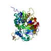
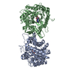
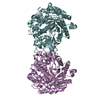
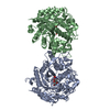
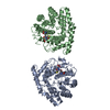
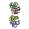
| Title | Crystal structure of the complex of Tdp1 with duplex DNA |
|---|
 Components Components | - DNA (5'-D(P*AP*AP*TP*GP*CP*GP*CP*AP*TP*TP*A)-3')
- Tyrosyl-DNA phosphodiesterase 1
|
|---|
 Keywords Keywords | HYDROLASE / protein-DNA complex / DNA repair / Nucleosidase / phosphotyrosine diesterase |
|---|
| Function / homology |  Function and homology information Function and homology information
3'-tyrosyl-DNA phosphodiesterase activity / Hydrolases; Acting on ester bonds; Phosphoric-diester hydrolases / single strand break repair / exonuclease activity / Nonhomologous End-Joining (NHEJ) / double-strand break repair / single-stranded DNA binding / double-stranded DNA binding / DNA repair / nucleoplasm ...3'-tyrosyl-DNA phosphodiesterase activity / Hydrolases; Acting on ester bonds; Phosphoric-diester hydrolases / single strand break repair / exonuclease activity / Nonhomologous End-Joining (NHEJ) / double-strand break repair / single-stranded DNA binding / double-stranded DNA binding / DNA repair / nucleoplasm / nucleus / plasma membrane / cytoplasmSimilarity search - Function Tyrosyl-DNA phosphodiesterase I / Tyrosyl-DNA phosphodiesterase / Endonuclease Chain A / Endonuclease; Chain A / 2-Layer Sandwich / Alpha BetaSimilarity search - Domain/homology |
|---|
| Biological species |  Homo sapiens (human) Homo sapiens (human)
synthetic construct (others) |
|---|
| Method |  X-RAY DIFFRACTION / SYNCHROTRON / MOLECULAR REPLACEMENT / molecular replacement / Resolution: 3.2 Å X-RAY DIFFRACTION / SYNCHROTRON / MOLECULAR REPLACEMENT / molecular replacement / Resolution: 3.2 Å |
|---|
| Model details | Tdp1 with product of nucleoside cleavage |
|---|
 Authors Authors | Richardson, J.M. / Ruksenaite, E. / Morris, E.R. |
|---|
 Citation Citation | Journal: Nat Commun / Year: 2018
Title: Structural basis for DNA 3'-end processing by human tyrosyl-DNA phosphodiesterase 1.
Authors: Flett, F.J. / Ruksenaite, E. / Armstrong, L.A. / Bharati, S. / Carloni, R. / Morris, E.R. / Mackay, C.L. / Interthal, H. / Richardson, J.M. |
|---|
| History | | Deposition | May 5, 2017 | Deposition site: PDBE / Processing site: PDBE |
|---|
| Revision 1.0 | Jan 10, 2018 | Provider: repository / Type: Initial release |
|---|
| Revision 1.1 | Jan 17, 2018 | Group: Database references / Category: citation / citation_author
Item: _citation.journal_volume / _citation.page_first ..._citation.journal_volume / _citation.page_first / _citation.page_last / _citation.pdbx_database_id_PubMed / _citation.title |
|---|
| Revision 1.2 | Oct 16, 2019 | Group: Data collection / Category: reflns_shell |
|---|
| Revision 1.3 | Jan 17, 2024 | Group: Data collection / Database references / Refinement description
Category: chem_comp_atom / chem_comp_bond ...chem_comp_atom / chem_comp_bond / database_2 / pdbx_initial_refinement_model
Item: _database_2.pdbx_DOI / _database_2.pdbx_database_accession |
|---|
|
|---|
 Movie
Movie Controller


 Open data
 Basic information
 Structure visualization
 Downloads & links
 Other downloads


 PDBj
PDBj


 Assembly


 Sample preparation
 / Beamline: I04 / Wavelength: 0.92819 Å
 Processing