 Movie
Movie Controller

[English] 日本語
 Yorodumi
- PDB-1v08: Crystal structure of the Zea maze beta-glucosidase-1 in complex w... -
+ Open data

- Basic information
| Entry | Database: PDB / ID: 1v08 | ||||||
|---|---|---|---|---|---|---|---|
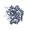
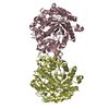
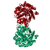
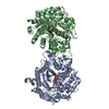
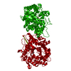
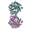
| Title | Crystal structure of the Zea maze beta-glucosidase-1 in complex with gluco-tetrazole | ||||||
 Components Components | BETA-GLUCOSIDASE | ||||||
 Keywords Keywords | HYDROLASE / BETA-GLUCOSIDASE / GLYCOSIDE HYDROLASE / DIMBOA-GLUCOSIDE / INHIBITOR / PEST DEFENSE / FAMILY GH1 / CHLOROPLAST / TRANSIT PEPTIDE | ||||||
| Function / homology |  Function and homology information Function and homology informationgalactosidase activity / fucosidase activity / xylanase activity / 4-hydroxy-7-methoxy-3-oxo-3,4-dihydro-2H-1,4-benzoxazin-2-yl glucoside beta-D-glucosidase / DIMBOA glucoside beta-D-glucosidase activity / cytokinin-activated signaling pathway / mannosidase activity / cellulose 1,4-beta-cellobiosidase activity / beta-glucosidase / beta-glucosidase activity ...galactosidase activity / fucosidase activity / xylanase activity / 4-hydroxy-7-methoxy-3-oxo-3,4-dihydro-2H-1,4-benzoxazin-2-yl glucoside beta-D-glucosidase / DIMBOA glucoside beta-D-glucosidase activity / cytokinin-activated signaling pathway / mannosidase activity / cellulose 1,4-beta-cellobiosidase activity / beta-glucosidase / beta-glucosidase activity / chloroplast / carbohydrate metabolic process Similarity search - Function | ||||||
| Biological species |  | ||||||
| Method |  X-RAY DIFFRACTION / SYNCHROTRON / MOLECULAR REPLACEMENT / Resolution: 1.9 Å X-RAY DIFFRACTION / SYNCHROTRON / MOLECULAR REPLACEMENT / Resolution: 1.9 Å | ||||||
 Authors Authors | Moriniere, J. / Verdoucq, L. / Bevan, D.R. / Esen, A. / Henrissat, B. / Czjzek, M. | ||||||
 Citation Citation | Journal: J.Biol.Chem. / Year: 2004 Title: Structural Determinants of Substrate Specificity in Family 1 Beta-Glucosidases: Novel Insights from the Crystal Structure of Sorghum Dhurrinase-1, a Plant Beta-Glucosidase with Strict ...Title: Structural Determinants of Substrate Specificity in Family 1 Beta-Glucosidases: Novel Insights from the Crystal Structure of Sorghum Dhurrinase-1, a Plant Beta-Glucosidase with Strict Specificity, in Complex with its Natural Substrate Authors: Verdoucq, L. / Moriniere, J. / Bevan, D.R. / Esen, A. / Vasella, A. / Henrissat, B. / Czjzek, M. | ||||||
| History |
| ||||||
| Remark 700 | SHEET DETERMINATION METHOD: DSSP THE SHEETS PRESENTED AS "AA BA" IN EACH CHAIN ON SHEET RECORDS ... SHEET DETERMINATION METHOD: DSSP THE SHEETS PRESENTED AS "AA BA" IN EACH CHAIN ON SHEET RECORDS BELOW IS ACTUALLY AN 9-STRANDED BARREL THIS IS REPRESENTED BY A 10-STRANDED SHEET IN WHICH THE FIRST AND LAST STRANDS ARE IDENTICAL. |
- Structure visualization
| Structure viewer | Molecule: MolmilJmol/JSmol |
|---|
- Downloads & links
-Download
| PDBx/mmCIF format | 1v08.cif.gz | 223.6 KB | Display | PDBx/mmCIF format |
|---|---|---|---|---|
| PDB format | pdb1v08.ent.gz | 180.5 KB | Display | PDB format |
| PDBx/mmJSON format | 1v08.json.gz | Tree view | PDBx/mmJSON format | |
| Others |  Other downloads Other downloads |
-Validation report
| Arichive directory | https://data.pdbj.org/pub/pdb/validation_reports/v0/1v08ftp://data.pdbj.org/pub/pdb/validation_reports/v0/1v08 | HTTPS FTP |
|---|
-Related structure data
| Related structure data |  1v02C  1v03C 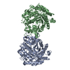 1e1eS S: Starting model for refinement C: citing same article ( |
|---|---|
| Similar structure data |


-Links
 PDBj
PDBj- Assembly
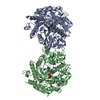
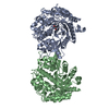
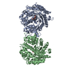
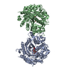
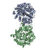
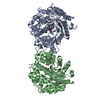
| Deposited unit | 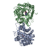
| ||||||||
|---|---|---|---|---|---|---|---|---|---|
| 1 |
| ||||||||
| Unit cell |
| ||||||||
| Noncrystallographic symmetry (NCS) | NCS oper: (Code: given Matrix: (-0.20717, -0.35231, -0.91267), Vector: |
-Components
| #1: Protein | Mass: 58458.422 Da / Num. of mol.: 2 / Mutation: YES Source method: isolated from a genetically manipulated source Source: (gene. exp.)  #2: Chemical | ChemComp-NTZ /   Mass: 202.168 Da / Num. of mol.: 4 / Source method: obtained synthetically / Formula: C6H10N4O4 Mass: 202.168 Da / Num. of mol.: 4 / Source method: obtained synthetically / Formula: C6H10N4O4#3: Water | ChemComp-HOH / |  Mass: 18.015 Da / Num. of mol.: 839 / Source method: isolated from a natural source / Formula: H2O Mass: 18.015 Da / Num. of mol.: 839 / Source method: isolated from a natural source / Formula: H2OCompound details | ENGINEERED MUTATION IN CHAIN A AND B, GLU 245 TO ASP CATALYTIC ACTIVITY: HYDROLYSIS OF TERMINAL, ...ENGINEERED | Has protein modification | Y | Sequence details | ASP A 191, SITE DIRECTED MUTATION | |
|---|
-Experimental details
-Experiment
| Experiment | Method: X-RAY DIFFRACTION / Number of used crystals: 1 |
|---|
- Sample preparation
| Crystal | Density Matthews: 2.89 Å3/Da / Density % sol: 57.5 % |
|---|---|
| Crystal grow | pH: 7.5 / Details: 22% PEG 4000, 5% ISOPROPANOL, 0.1 M HEPES PH 7.5 |
-Data collection
| Diffraction | Mean temperature: 100 K |
|---|---|
| Diffraction source | Source: SYNCHROTRON / Site: ESRF  / Beamline: ID14-2 / Wavelength: 0.934 / Beamline: ID14-2 / Wavelength: 0.934 |
| Detector | Date: Feb 15, 2001 |
| Radiation | Protocol: SINGLE WAVELENGTH / Monochromatic (M) / Laue (L): M / Scattering type: x-ray |
| Radiation wavelength | Wavelength: 0.934 Å / Relative weight: 1 |
| Reflection | Resolution: 1.95→29.8 Å / Num. obs: 92037 / % possible obs: 96.6 % / Redundancy: 3.3 % / Rmerge(I) obs: 0.072 / Net I/σ(I): 6.9 |
| Reflection shell | Resolution: 1.95→2.01 Å / Redundancy: 3.2 % / Rmerge(I) obs: 0.456 / Mean I/σ(I) obs: 1.5 / % possible all: 94 |
- Processing
| Software |
| ||||||||||||||||||||||||||||||||||||||||||||||||||||||||||||||||||||||||||||||||||||||||||||||||||||||||||||||||||||||||||||||||||||||||||||||||||||||||||||||||||||||||||||||||||||||
|---|---|---|---|---|---|---|---|---|---|---|---|---|---|---|---|---|---|---|---|---|---|---|---|---|---|---|---|---|---|---|---|---|---|---|---|---|---|---|---|---|---|---|---|---|---|---|---|---|---|---|---|---|---|---|---|---|---|---|---|---|---|---|---|---|---|---|---|---|---|---|---|---|---|---|---|---|---|---|---|---|---|---|---|---|---|---|---|---|---|---|---|---|---|---|---|---|---|---|---|---|---|---|---|---|---|---|---|---|---|---|---|---|---|---|---|---|---|---|---|---|---|---|---|---|---|---|---|---|---|---|---|---|---|---|---|---|---|---|---|---|---|---|---|---|---|---|---|---|---|---|---|---|---|---|---|---|---|---|---|---|---|---|---|---|---|---|---|---|---|---|---|---|---|---|---|---|---|---|---|---|---|---|---|
| Refinement | Method to determine structure: MOLECULAR REPLACEMENT Starting model: PDB ENTRY 1E1E Resolution: 1.9→29.75 Å / Cor.coef. Fo:Fc: 0.962 / Cor.coef. Fo:Fc free: 0.949 / SU B: 2.826 / SU ML: 0.078 / Cross valid method: THROUGHOUT / ESU R: 0.138 / ESU R Free: 0.126 / Stereochemistry target values: MAXIMUM LIKELIHOOD / Details: HYDROGENS HAVE BEEN ADDED IN THE RIDING POSITIONS
| ||||||||||||||||||||||||||||||||||||||||||||||||||||||||||||||||||||||||||||||||||||||||||||||||||||||||||||||||||||||||||||||||||||||||||||||||||||||||||||||||||||||||||||||||||||||
| Solvent computation | Ion probe radii: 0.8 Å / Shrinkage radii: 0.8 Å / VDW probe radii: 1.4 Å / Solvent model: BABINET MODEL WITH MASK | ||||||||||||||||||||||||||||||||||||||||||||||||||||||||||||||||||||||||||||||||||||||||||||||||||||||||||||||||||||||||||||||||||||||||||||||||||||||||||||||||||||||||||||||||||||||
| Displacement parameters | Biso mean: 22.38 Å2
| ||||||||||||||||||||||||||||||||||||||||||||||||||||||||||||||||||||||||||||||||||||||||||||||||||||||||||||||||||||||||||||||||||||||||||||||||||||||||||||||||||||||||||||||||||||||
| Refinement step | Cycle: LAST / Resolution: 1.9→29.75 Å
| ||||||||||||||||||||||||||||||||||||||||||||||||||||||||||||||||||||||||||||||||||||||||||||||||||||||||||||||||||||||||||||||||||||||||||||||||||||||||||||||||||||||||||||||||||||||
| Refine LS restraints |
|