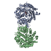
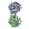
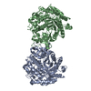
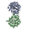
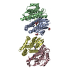
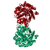
[English] 日本語
Yorodumi
- PDB-1v03: Crystal structure of the Sorghum bicolor dhurrinase 1 -

+
Open data


ID or keywords:

Loading...

-
Basic information

Entry
Database: PDB / ID: 1v03
TitleCrystal structure of the Sorghum bicolor dhurrinase 1
ComponentsDHURRINASE
KeywordsHYDROLASE / BETA-GLUCOSIDASE / DHURRIN HYDROLYSIS / PEST DEFENSE / FAMILY GH1
Function / homology
Function and homology information


beta-glucosidase activity / chloroplast / carbohydrate metabolic process
Similarity search - Function
Glycoside hydrolase family 1, active site / Glycosyl hydrolases family 1 active site. / Glycosyl hydrolases family 1, N-terminal conserved site / Glycosyl hydrolases family 1 N-terminal signature. / Glycosyl hydrolase family 1 / Glycoside hydrolase family 1 / Glycosidases / Glycoside hydrolase superfamily / TIM Barrel / Alpha-Beta Barrel / Alpha Beta
Similarity search - Domain/homology
beta-D-glucopyranose / ACETONITRILE / PHENOL / Dhurrinase
Similarity search - Component
Biological speciesSORGHUM BICOLOR MILO (sorghum)
MethodX-RAY DIFFRACTION / SYNCHROTRON / MOLECULAR REPLACEMENT / Resolution: 2 Å
AuthorsMoriniere, J. / Verdoucq, L. / Bevan, D.R. / Esen, A. / Henrissat, B. / Czjzek, M.
CitationJournal: J.Biol.Chem. / Year: 2004
Title: Structural Determinants of Substrate Specificity in Family 1 Beta-Glucosidases: Novel Insights from the Crystal Structure of Sorghum Dhurrinase-1, a Plant Beta-Glucosidase with Strict ...Title: Structural Determinants of Substrate Specificity in Family 1 Beta-Glucosidases: Novel Insights from the Crystal Structure of Sorghum Dhurrinase-1, a Plant Beta-Glucosidase with Strict Specificity, in Complex with its Natural Substrate
Authors: Verdoucq, L. / Moriniere, J. / Bevan, D.R. / Esen, A. / Vasella, A. / Henrissat, B. / Czjzek, M.
History
DepositionMar 22, 2004Deposition site: PDBE / Processing site: PDBE
Revision 1.0May 20, 2004Provider: repository / Type: Initial release
Revision 1.1May 7, 2011Group: Version format compliance
Revision 1.2Jul 13, 2011Group: Version format compliance
Revision 1.3Jul 29, 2020Group: Data collection / Derived calculations ...Data collection / Derived calculations / Other / Structure summary
Category: chem_comp / entity ...chem_comp / entity / pdbx_chem_comp_identifier / pdbx_database_status / pdbx_entity_nonpoly / struct_conn / struct_site / struct_site_gen
Item: _chem_comp.name / _chem_comp.type ..._chem_comp.name / _chem_comp.type / _entity.pdbx_description / _pdbx_database_status.status_code_sf / _pdbx_entity_nonpoly.name / _struct_conn.pdbx_leaving_atom_flag
Description: Carbohydrate remediation / Provider: repository / Type: Remediation
Revision 1.4Dec 13, 2023Group: Data collection / Database references ...Data collection / Database references / Refinement description / Structure summary
Category: chem_comp / chem_comp_atom ...chem_comp / chem_comp_atom / chem_comp_bond / database_2 / pdbx_initial_refinement_model
Item: _chem_comp.pdbx_synonyms / _database_2.pdbx_DOI / _database_2.pdbx_database_accession
Remark 700 SHEET DETERMINATION METHOD: DSSP THE SHEETS PRESENTED AS "AA" IN EACH CHAIN ON SHEET RECORDS BELOW ... SHEET DETERMINATION METHOD: DSSP THE SHEETS PRESENTED AS "AA" IN EACH CHAIN ON SHEET RECORDS BELOW IS ACTUALLY AN 9-STRANDED BARREL THIS IS REPRESENTED BY A 10-STRANDED SHEET IN WHICH THE FIRST AND LAST STRANDS ARE IDENTICAL.

-
Structure visualization

Structure viewerMolecule:
MolmilJmol/JSmol

Downloads & links

-
Assembly

Deposited unit
A: DHURRINASE
hetero molecules


Theoretical massNumber of molelcules
Total (without water)64,1474
Polymers63,8321
Non-polymers3153
Water10,809600
1
A: DHURRINASE
hetero molecules

A: DHURRINASE
hetero molecules


Theoretical massNumber of molelcules
Total (without water)128,2948
Polymers127,6632
Non-polymers6316
Water362
TypeNameSymmetry operationNumber
identity operation1_555x,y,z1
crystal symmetry operation19_555-x+3/4,-z+3/4,-y+3/41
MethodPQS
Unit cell
Length a, b, c (Å)195.490, 195.490, 195.490
Angle α, β, γ (deg.)90.00, 90.00, 90.00
Int Tables number213
Space group name H-MP4132
Components on special symmetry positions
IDModelComponents
11A-2033-

HOH

-
Components

#1: Protein DHURRINASE / DHURRINASE-1


Mass: 63831.613 Da / Num. of mol.: 1 / Mutation: YES
Source method: isolated from a genetically manipulated source
Source: (gene. exp.) SORGHUM BICOLOR MILO (sorghum) / Production host: ESCHERICHIA COLI (E. coli) / References: UniProt: Q41290, beta-glucosidase
#2: Sugar ChemComp-BGC / beta-D-glucopyranose / beta-D-glucose / D-glucose / glucose / Glucose


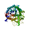
Type: D-saccharide, beta linking / Mass: 180.156 Da / Num. of mol.: 1
Source method: isolated from a genetically manipulated source
Formula: C6H12O6
IdentifierTypeProgram
DGlcpbCONDENSED IUPAC CARBOHYDRATE SYMBOLGMML 1.0
b-D-glucopyranoseCOMMON NAMEGMML 1.0
b-D-GlcpIUPAC CARBOHYDRATE SYMBOLPDB-CARE 1.0
GlcSNFG CARBOHYDRATE SYMBOLGMML 1.0
#3: Chemical ChemComp-CCN / ACETONITRILE / Acetonitrile


Mass: 41.052 Da / Num. of mol.: 1 / Source method: obtained synthetically / Formula: C2H3N
#4: Chemical ChemComp-IPH / PHENOL / Phenol


Mass: 94.111 Da / Num. of mol.: 1 / Source method: obtained synthetically / Formula: C6H6O
#5: Water ChemComp-HOH / water / Water


Mass: 18.015 Da / Num. of mol.: 600 / Source method: isolated from a natural source / Formula: H2O
Compound detailsENGINEERED RESIDUE CHAIN A: GLU 240 ASP

-
Experimental details

-
Experiment

ExperimentMethod: X-RAY DIFFRACTION / Number of used crystals: 1

-
Sample preparation

CrystalDensity Matthews: 5.98 Å3/Da / Density % sol: 79 %
Crystal growpH: 7.5 / Details: 2.0 M NH4(SO4)2, 0.1 M HEPERS PH 7.5, 50 MM NACL

-
Data collection

DiffractionMean temperature: 100 K
Diffraction sourceSource: SYNCHROTRON / Site: ESRF / Beamline: ID14-2 / Wavelength: 0.934
DetectorDate: Apr 15, 2003
RadiationProtocol: SINGLE WAVELENGTH / Monochromatic (M) / Laue (L): M / Scattering type: x-ray
Radiation wavelengthWavelength: 0.934 Å / Relative weight: 1
ReflectionResolution: 2→39 Å / Num. obs: 85811 / % possible obs: 99.8 % / Redundancy: 7.9 % / Rmerge(I) obs: 0.073 / Net I/σ(I): 6.8
Reflection shellResolution: 2→2.07 Å / Redundancy: 7 % / Rmerge(I) obs: 0.328 / Mean I/σ(I) obs: 2.2 / % possible all: 99.7

-
Processing

Software
NameVersionClassification
REFMAC5.1.24refinement
DENZOdata reduction
SCALAdata scaling
AMoREphasing
RefinementMethod to determine structure: MOLECULAR REPLACEMENT
Starting model: PDB ENTRY 1E1E
Resolution: 2→141.42 Å / Cor.coef. Fo:Fc: 0.954 / Cor.coef. Fo:Fc free: 0.941 / SU B: 2.452 / SU ML: 0.068 / Cross valid method: THROUGHOUT / ESU R: 0.102 / ESU R Free: 0.102 / Stereochemistry target values: MAXIMUM LIKELIHOOD
RfactorNum. reflection% reflectionSelection details
Rfree0.213 4294 5 %RANDOM
Rwork0.187 ---
obs0.189 81445 99.8 %-
Solvent computationIon probe radii: 0.8 Å / Shrinkage radii: 0.8 Å / VDW probe radii: 1.4 Å / Solvent model: BABINET MODEL WITH MASK
Displacement parametersBiso mean: 28.89 Å2
Refinement stepCycle: LAST / Resolution: 2→141.42 Å
ProteinNucleic acidLigandSolventTotal
Num. atoms3898 0 22 600 4520
Refine LS restraints
Refine-IDTypeDev idealDev ideal targetNumber
X-RAY DIFFRACTIONr_bond_refined_d0.0170.0214037
X-RAY DIFFRACTIONr_bond_other_d
X-RAY DIFFRACTIONr_angle_refined_deg1.5291.9415482
X-RAY DIFFRACTIONr_angle_other_deg
X-RAY DIFFRACTIONr_dihedral_angle_1_deg5.8495486
X-RAY DIFFRACTIONr_dihedral_angle_2_deg
X-RAY DIFFRACTIONr_dihedral_angle_3_deg
X-RAY DIFFRACTIONr_dihedral_angle_4_deg
X-RAY DIFFRACTIONr_chiral_restr0.1120.2562
X-RAY DIFFRACTIONr_gen_planes_refined0.0070.023166
X-RAY DIFFRACTIONr_gen_planes_other
X-RAY DIFFRACTIONr_nbd_refined0.2430.32125
X-RAY DIFFRACTIONr_nbd_other
X-RAY DIFFRACTIONr_nbtor_refined
X-RAY DIFFRACTIONr_nbtor_other
X-RAY DIFFRACTIONr_xyhbond_nbd_refined0.2050.5840
X-RAY DIFFRACTIONr_xyhbond_nbd_other
X-RAY DIFFRACTIONr_metal_ion_refined
X-RAY DIFFRACTIONr_metal_ion_other
X-RAY DIFFRACTIONr_symmetry_vdw_refined0.1440.330
X-RAY DIFFRACTIONr_symmetry_vdw_other
X-RAY DIFFRACTIONr_symmetry_hbond_refined0.2530.548
X-RAY DIFFRACTIONr_symmetry_hbond_other
X-RAY DIFFRACTIONr_symmetry_metal_ion_refined
X-RAY DIFFRACTIONr_symmetry_metal_ion_other
X-RAY DIFFRACTIONr_mcbond_it1.42222420
X-RAY DIFFRACTIONr_mcbond_other
X-RAY DIFFRACTIONr_mcangle_it2.33233898
X-RAY DIFFRACTIONr_mcangle_other
X-RAY DIFFRACTIONr_scbond_it1.70221617
X-RAY DIFFRACTIONr_scbond_other
X-RAY DIFFRACTIONr_scangle_it2.5731584
X-RAY DIFFRACTIONr_scangle_other
X-RAY DIFFRACTIONr_long_range_B_refined
X-RAY DIFFRACTIONr_long_range_B_other
X-RAY DIFFRACTIONr_rigid_bond_restr
X-RAY DIFFRACTIONr_sphericity_free
X-RAY DIFFRACTIONr_sphericity_bonded
LS refinement shellResolution: 2→2.05 Å / Total num. of bins used: 20 /
RfactorNum. reflection
Rfree0.293 310
Rwork0.249 5934

+
About Yorodumi

-
News

-
Feb 9, 2022. New format data for meta-information of EMDB entries

New format data for meta-information of EMDB entries

  • Version 3 of the EMDB header file is now the official format.
  • The previous official version 1.9 will be removed from the archive.

Related info.:EMDB header

External links:wwPDB to switch to version 3 of the EMDB data model

-
Aug 12, 2020. Covid-19 info

Covid-19 info

URL: https://pdbj.org/emnavi/covid19.php

New page: Covid-19 featured information page in EM Navigator.

Related info.:Covid-19 info / Mar 5, 2020. Novel coronavirus structure data

+
Mar 5, 2020. Novel coronavirus structure data

Novel coronavirus structure data

Related info.:Yorodumi Speices / Aug 12, 2020. Covid-19 info

External links:COVID-19 featured content - PDBj / Molecule of the Month (242):Coronavirus Proteases

+
Jan 31, 2019. EMDB accession codes are about to change! (news from PDBe EMDB page)

EMDB accession codes are about to change! (news from PDBe EMDB page)

  • The allocation of 4 digits for EMDB accession codes will soon come to an end. Whilst these codes will remain in use, new EMDB accession codes will include an additional digit and will expand incrementally as the available range of codes is exhausted. The current 4-digit format prefixed with “EMD-” (i.e. EMD-XXXX) will advance to a 5-digit format (i.e. EMD-XXXXX), and so on. It is currently estimated that the 4-digit codes will be depleted around Spring 2019, at which point the 5-digit format will come into force.
  • The EM Navigator/Yorodumi systems omit the EMD- prefix.

Related info.:Q: What is EMD? / ID/Accession-code notation in Yorodumi/EM Navigator

External links:EMDB Accession Codes are Changing Soon! / Contact to PDBj

+
Jul 12, 2017. Major update of PDB

Major update of PDB

  • wwPDB released updated PDB data conforming to the new PDBx/mmCIF dictionary.
  • This is a major update changing the version number from 4 to 5, and with Remediation, in which all the entries are updated.
  • In this update, many items about electron microscopy experimental information are reorganized (e.g. em_software).
  • Now, EM Navigator and Yorodumi are based on the updated data.

External links:wwPDB Remediation / Enriched Model Files Conforming to OneDep Data Standards Now Available in the PDB FTP Archive

-
Yorodumi

Thousand views of thousand structures

  • Yorodumi is a browser for structure data from EMDB, PDB, SASBDB, etc.
  • This page is also the successor to EM Navigator detail page, and also detail information page/front-end page for Omokage search.
  • The word "yorodu" (or yorozu) is an old Japanese word meaning "ten thousand". "mi" (miru) is to see.

Related info.:EMDB / PDB / SASBDB / Comparison of 3 databanks / Yorodumi Search / Aug 31, 2016. New EM Navigator & Yorodumi / Yorodumi Papers / Jmol/JSmol / Function and homology information / Changes in new EM Navigator and Yorodumi

Read more