 Movie
Movie Controller


+ Open data

- Basic information
| Entry | Database: PDB / ID: 1e1e | ||||||
|---|---|---|---|---|---|---|---|
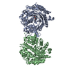
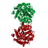
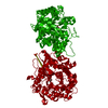
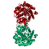
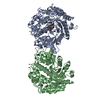
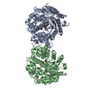
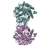
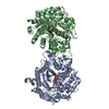
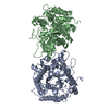
| Title | Crystal structure of a Monocot (Maize ZMGlu1) beta-glucosidase | ||||||
 Components Components | BETA-GLUCOSIDASE | ||||||
 Keywords Keywords | HYDROLASE / GLYCOSIDE HYDROLASE / BETA-GLUCOSIDASE / FAMILY 1 / RETENTION OF THE ANOMERIC CONFIGURATION | ||||||
| Function / homology |  Function and homology information Function and homology informationgalactosidase activity / fucosidase activity / xylanase activity / 4-hydroxy-7-methoxy-3-oxo-3,4-dihydro-2H-1,4-benzoxazin-2-yl glucoside beta-D-glucosidase / DIMBOA glucoside beta-D-glucosidase activity / cytokinin-activated signaling pathway / mannosidase activity / cellulose 1,4-beta-cellobiosidase activity / beta-glucosidase / beta-glucosidase activity ...galactosidase activity / fucosidase activity / xylanase activity / 4-hydroxy-7-methoxy-3-oxo-3,4-dihydro-2H-1,4-benzoxazin-2-yl glucoside beta-D-glucosidase / DIMBOA glucoside beta-D-glucosidase activity / cytokinin-activated signaling pathway / mannosidase activity / cellulose 1,4-beta-cellobiosidase activity / beta-glucosidase / beta-glucosidase activity / chloroplast / carbohydrate metabolic process Similarity search - Function | ||||||
| Biological species |  | ||||||
| Method |  X-RAY DIFFRACTION / SYNCHROTRON / MOLECULAR REPLACEMENT / Resolution: 2.5 Å X-RAY DIFFRACTION / SYNCHROTRON / MOLECULAR REPLACEMENT / Resolution: 2.5 Å | ||||||
 Authors Authors | Czjzek, M. / Cicek, M. / Bevan, D.R. / Henrissat, B. / Esen, A. | ||||||
 Citation Citation | Journal: Biochem.J. / Year: 2001 Title: Crystal Structure of a Monocotyledon (Maize Zmglu1) Beta-Glucosidase and a Model of its Complex with P-Nitrophenyl Beta-D-Thioglucoside Authors: Czjzek, M. / Cicek, M. / Zamboni, V. / Burmeister, W.P. / Bevan, D.R. / Henrissat, B. / Esen, A. #1: Journal: Plant Physiol. / Year: 1996Title: Nucleotide Sequence of a Cdna Corresponding to a Second Beta-Glucosidase Gene in Maize Authors: Bandaranayake, H. / Esen, A. | ||||||
| History |
|
- Structure visualization
| Structure viewer | Molecule: MolmilJmol/JSmol |
|---|
- Downloads & links
-Download
| PDBx/mmCIF format | 1e1e.cif.gz | 204.3 KB | Display | PDBx/mmCIF format |
|---|---|---|---|---|
| PDB format | pdb1e1e.ent.gz | 166.4 KB | Display | PDB format |
| PDBx/mmJSON format | 1e1e.json.gz | Tree view | PDBx/mmJSON format | |
| Others |  Other downloads Other downloads |
-Validation report
| Arichive directory | https://data.pdbj.org/pub/pdb/validation_reports/e1/1e1eftp://data.pdbj.org/pub/pdb/validation_reports/e1/1e1e | HTTPS FTP |
|---|
-Related structure data
| Related structure data | 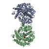 1e1fC  1cbgS S: Starting model for refinement C: citing same article ( |
|---|---|
| Similar structure data |


-Links
 PDBj
PDBj- Assembly
| Deposited unit | 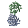
| ||||||||
|---|---|---|---|---|---|---|---|---|---|
| 1 |
| ||||||||
| Unit cell |
| ||||||||
| Noncrystallographic symmetry (NCS) | NCS oper: (Code: given Matrix: (0.88564, 0.35057, 0.30456), Vector: |
-Components
| #1: Protein | Mass: 58472.445 Da / Num. of mol.: 2 Source method: isolated from a genetically manipulated source Source: (gene. exp.)  #2: Water | ChemComp-HOH / |  Mass: 18.015 Da / Num. of mol.: 331 / Source method: isolated from a natural source / Formula: H2O Mass: 18.015 Da / Num. of mol.: 331 / Source method: isolated from a natural source / Formula: H2OHas protein modification | Y | |
|---|
-Experimental details
-Experiment
| Experiment | Method: X-RAY DIFFRACTION / Number of used crystals: 1 |
|---|
- Sample preparation
| Crystal | Density Matthews: 2.5 Å3/Da / Density % sol: 50.86 % | ||||||||||||||||||||||||||||||
|---|---|---|---|---|---|---|---|---|---|---|---|---|---|---|---|---|---|---|---|---|---|---|---|---|---|---|---|---|---|---|---|
| Crystal grow | pH: 5.6 / Details: 0.2 M AMMONIUM SULFATE, pH 5.60 | ||||||||||||||||||||||||||||||
| Crystal grow | *PLUS pH: 7 / Method: vapor diffusion, hanging drop | ||||||||||||||||||||||||||||||
| Components of the solutions | *PLUS
|
-Data collection
| Diffraction | Mean temperature: 100 K |
|---|---|
| Diffraction source | Source: SYNCHROTRON / Site: ESRF  / Beamline: ID14-3 / Wavelength: 0.93 / Beamline: ID14-3 / Wavelength: 0.93 |
| Detector | Date: Jan 15, 1999 |
| Radiation | Protocol: SINGLE WAVELENGTH / Monochromatic (M) / Laue (L): M / Scattering type: x-ray |
| Radiation wavelength | Wavelength: 0.93 Å / Relative weight: 1 |
| Reflection | Resolution: 2.42→29.1 Å / Num. obs: 549492 / % possible obs: 99 % / Redundancy: 3.6 % / Rmerge(I) obs: 0.074 / Rsym value: 0.11 / Net I/σ(I): 6.4 |
| Reflection shell | Resolution: 2.34→2.42 Å / Redundancy: 3.6 % / Rmerge(I) obs: 0.074 / Mean I/σ(I) obs: 2.3 / Rsym value: 0.207 / % possible all: 99 |
| Reflection | *PLUS Lowest resolution: 27 Å / Num. obs: 42762 / % possible obs: 99.9 % / Num. measured all: 153615 / Rmerge(I) obs: 0.093 |
| Reflection shell | *PLUS % possible obs: 99.9 % / Rmerge(I) obs: 0.276 |
- Processing
| Software |
| ||||||||||||||||||||||||||||||||||||||||||||||||||||||||||||
|---|---|---|---|---|---|---|---|---|---|---|---|---|---|---|---|---|---|---|---|---|---|---|---|---|---|---|---|---|---|---|---|---|---|---|---|---|---|---|---|---|---|---|---|---|---|---|---|---|---|---|---|---|---|---|---|---|---|---|---|---|---|
| Refinement | Method to determine structure: MOLECULAR REPLACEMENT Starting model: PDB ENTRY 1CBG Resolution: 2.5→27 Å / Data cutoff high absF: 10000000 / Cross valid method: THROUGHOUT / σ(F): 0.1
| ||||||||||||||||||||||||||||||||||||||||||||||||||||||||||||
| Solvent computation | Solvent model: FLAT MODEL / Bsol: 48.69 Å2 / ksol: 0.325 e/Å3 | ||||||||||||||||||||||||||||||||||||||||||||||||||||||||||||
| Displacement parameters | Biso mean: 50.39 Å2 | ||||||||||||||||||||||||||||||||||||||||||||||||||||||||||||
| Refine analyze |
| ||||||||||||||||||||||||||||||||||||||||||||||||||||||||||||
| Refinement step | Cycle: LAST / Resolution: 2.5→27 Å
| ||||||||||||||||||||||||||||||||||||||||||||||||||||||||||||
| Refine LS restraints |
| ||||||||||||||||||||||||||||||||||||||||||||||||||||||||||||
| Refine LS restraints NCS | Rms dev position: 0.362 Å / Weight position: 10 | ||||||||||||||||||||||||||||||||||||||||||||||||||||||||||||
| LS refinement shell | Resolution: 2.5→2.59 Å / Total num. of bins used: 10
| ||||||||||||||||||||||||||||||||||||||||||||||||||||||||||||
| Xplor file | Serial no: 1 / Param file: PROTEIN_REP.PARAM / Topol file: PROTEIN.TOP | ||||||||||||||||||||||||||||||||||||||||||||||||||||||||||||
| Software | *PLUS Name: CNS / Version: 1 / Classification: refinement | ||||||||||||||||||||||||||||||||||||||||||||||||||||||||||||
| Refinement | *PLUS Rfactor obs: 0.24 / Rfactor Rfree: 0.283 | ||||||||||||||||||||||||||||||||||||||||||||||||||||||||||||
| Solvent computation | *PLUS | ||||||||||||||||||||||||||||||||||||||||||||||||||||||||||||
| Displacement parameters | *PLUS | ||||||||||||||||||||||||||||||||||||||||||||||||||||||||||||
| Refine LS restraints | *PLUS
|
