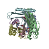
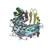
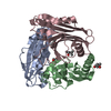
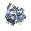
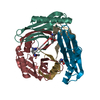
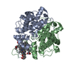
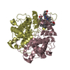
#1: Journal: J.Biol.Inorg.Chem. / Year: 1997 Title: A Preliminary Analysis of the Three Dimensional Structure of Dimeric Di-Haem Split-Soret Cytochrome C from Desulfovibrio Desulfuricans Atcc 27774 at 2.5 A Resolution Using the MAD Phasing ...Title: A Preliminary Analysis of the Three Dimensional Structure of Dimeric Di-Haem Split-Soret Cytochrome C from Desulfovibrio Desulfuricans Atcc 27774 at 2.5 A Resolution Using the MAD Phasing Method: A Novel Cytochrome Fold with a Stacked Haem Arrangement Authors: Matias, P.M. / Morais, J. / Coelho, A.V. / Meijers, R. / Gonzalez, A. / Thompson, A.W. / Sieker, L. / Legall, J. / Carrondo, M.A.
Mass: 18.015 Da / Num. of mol.: 188 / Source method: isolated from a natural source / Formula: H2O
Has protein modification
Y
Sequence details
GENBANK ENTRY AF465622 IS ON HOLD PENDING MANUSCRIPT ACCEPTANCE FOR PUBLICATION. SEQADV RECORDS ...GENBANK ENTRY AF465622 IS ON HOLD PENDING MANUSCRIPT ACCEPTANCE FOR PUBLICATION. SEQADV RECORDS BELOW ARE DUE TO ERRORS IN SWS P81040 THAT WERE CORRECTED IN AF465622
-
Experimental details
-
Experiment
Experiment
Method: X-RAY DIFFRACTION / Number of used crystals: 1
-
Sample preparation
Crystal
Density Matthews: 2.59 Å3/Da / Density % sol: 53 %
Crystal grow
pH: 5 Details: PROTEIN WAS CRYSTALLIZED FROM A SOLUTION CONTAINING 0.1-0.2 % (W/V) AGAROSE IN 12-15 % PEG 8K, 0.1M SODIUM ACETATE BUFFER PH 5.0
Monochromator: SI (111) / Protocol: MAD / Monochromatic (M) / Laue (L): M / Scattering type: x-ray
Radiation wavelength
Wavelength: 1.7401 Å / Relative weight: 1
Reflection
Resolution: 2.5→20 Å / Num. obs: 34228 / % possible obs: 93 % / Redundancy: 6.2 % / Rmerge(I) obs: 0.038 / Net I/σ(I): 9.7
Reflection shell
Resolution: 2.5→2.56 Å / Rmerge(I) obs: 0.074 / Mean I/σ(I) obs: 9.7 / % possible all: 86
-
Processing
Software
Name
Version
Classification
DENZO
datareduction
SCALEPACK
datascaling
MLPHARE
phasing
DM
phasing
X-PLOR
3.1
refinement
Refinement
Method to determine structure: MAD / Resolution: 2.5→20 Å / Data cutoff high absF: 100000 / Data cutoff low absF: 0.1 / Isotropic thermal model: RESTRAINED / Cross valid method: THROUGHOUT / σ(F): 2 / Details: FIRST 7 RESIDUES NOT VISIBLE IN ELECTRON DENSITY.
Rfactor
Num. reflection
% reflection
Selection details
Rfree
0.264
1681
5 %
RANDOM
Rwork
0.199
-
-
-
obs
0.199
34127
92.8 %
-
Displacement parameters
Biso mean: 35.04 Å2
Refine analyze
Luzzati d res low obs: 10 Å / Luzzati sigma a obs: 0.35 Å
Refinement step
Cycle: LAST / Resolution: 2.5→20 Å
Protein
Nucleic acid
Ligand
Solvent
Total
Num. atoms
7384
0
344
188
7916
Refine LS restraints
Refine-ID
Type
Dev ideal
X-RAY DIFFRACTION
x_bond_d
0.013
X-RAY DIFFRACTION
x_bond_d_na
X-RAY DIFFRACTION
x_bond_d_prot
X-RAY DIFFRACTION
x_angle_d
X-RAY DIFFRACTION
x_angle_d_na
X-RAY DIFFRACTION
x_angle_d_prot
X-RAY DIFFRACTION
x_angle_deg
1.65
X-RAY DIFFRACTION
x_angle_deg_na
X-RAY DIFFRACTION
x_angle_deg_prot
X-RAY DIFFRACTION
x_dihedral_angle_d
24.5
X-RAY DIFFRACTION
x_dihedral_angle_d_na
X-RAY DIFFRACTION
x_dihedral_angle_d_prot
X-RAY DIFFRACTION
x_improper_angle_d
1.76
X-RAY DIFFRACTION
x_improper_angle_d_na
X-RAY DIFFRACTION
x_improper_angle_d_prot
X-RAY DIFFRACTION
x_mcbond_it
X-RAY DIFFRACTION
x_mcangle_it
X-RAY DIFFRACTION
x_scbond_it
X-RAY DIFFRACTION
x_scangle_it
Xplor file
Refine-ID
Serial no
Param file
Topol file
X-RAY DIFFRACTION
1
PARHCSDX.PRO
TOPHCSDX.PRO
X-RAY DIFFRACTION
2
PARAM19X.HEME
TOPH19X.HEME
Refine LS restraints
*PLUS
Refine-ID
Type
Dev ideal
X-RAY DIFFRACTION
x_dihedral_angle_d
X-RAY DIFFRACTION
x_dihedral_angle_deg
24.5
X-RAY DIFFRACTION
x_improper_angle_d
X-RAY DIFFRACTION
x_improper_angle_deg
1.76
+
About Yorodumi
-
News
-
Feb 9, 2022. New format data for meta-information of EMDB entries
New format data for meta-information of EMDB entries
Version 3 of the EMDB header file is now the official format.
The previous official version 1.9 will be removed from the archive.
In the structure databanks used in Yorodumi, some data are registered as the other names, "COVID-19 virus" and "2019-nCoV". Here are the details of the virus and the list of structure data.
Jan 31, 2019. EMDB accession codes are about to change! (news from PDBe EMDB page)
EMDB accession codes are about to change! (news from PDBe EMDB page)
The allocation of 4 digits for EMDB accession codes will soon come to an end. Whilst these codes will remain in use, new EMDB accession codes will include an additional digit and will expand incrementally as the available range of codes is exhausted. The current 4-digit format prefixed with “EMD-” (i.e. EMD-XXXX) will advance to a 5-digit format (i.e. EMD-XXXXX), and so on. It is currently estimated that the 4-digit codes will be depleted around Spring 2019, at which point the 5-digit format will come into force.
The EM Navigator/Yorodumi systems omit the EMD- prefix.
Related info.:Q: What is EMD? / ID/Accession-code notation in Yorodumi/EM Navigator
Yorodumi is a browser for structure data from EMDB, PDB, SASBDB, etc.
This page is also the successor to EM Navigator detail page, and also detail information page/front-end page for Omokage search.
The word "yorodu" (or yorozu) is an old Japanese word meaning "ten thousand". "mi" (miru) is to see.
Related info.:EMDB / PDB / SASBDB / Comparison of 3 databanks / Yorodumi Search / Aug 31, 2016. New EM Navigator & Yorodumi / Yorodumi Papers / Jmol/JSmol / Function and homology information / Changes in new EM Navigator and Yorodumi
 Movie
Movie Controller

 Yorodumi
 Open data
 Basic information
 Components
 Keywords
 Function and homology information
 DESULFOVIBRIO DESULFURICANS (bacteria)
 X-RAY DIFFRACTION /
  Authors
 Citation
 Structure visualization
 Downloads & links
 Other downloads
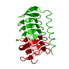


 PDBj
PDBj


 Assembly

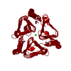
 Mass: 618.503 Da / Num. of mol.: 8 / Source method: obtained synthetically / Formula: C34H34FeN4O4
 Mass: 18.015 Da / Num. of mol.: 188 / Source method: isolated from a natural source / Formula: H2O
 Sample preparation
 / Beamline: BM14 / Wavelength: 1.7401
  Processing