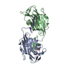
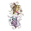
 Movie
Movie Controller


+ Open data

- Basic information
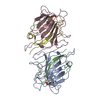
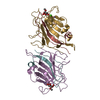
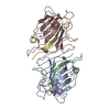
| Entry | Database: PDB / ID: 5nrq | ||||||
|---|---|---|---|---|---|---|---|
| Title | Mtb TMK crystal structure in complex with compound 33 | ||||||
 Components Components | Thymidylate kinase | ||||||
 Keywords Keywords | TRANSFERASE / Thymidylate kinase / Nucleotide Binding / inhibitor | ||||||
| Function / homology |  Function and homology information Function and homology informationTMP metabolic process / dTMP kinase / dUDP biosynthetic process / dTDP biosynthetic process / dTMP kinase activity / dTTP biosynthetic process / GTP binding / magnesium ion binding / protein homodimerization activity / ATP binding ...TMP metabolic process / dTMP kinase / dUDP biosynthetic process / dTDP biosynthetic process / dTMP kinase activity / dTTP biosynthetic process / GTP binding / magnesium ion binding / protein homodimerization activity / ATP binding / cytosol / cytoplasm Similarity search - Function | ||||||
| Biological species |   Mycobacterium tuberculosis (bacteria) Mycobacterium tuberculosis (bacteria) | ||||||
| Method |  X-RAY DIFFRACTION / SYNCHROTRON / MOLECULAR REPLACEMENT / Resolution: 2.1 Å X-RAY DIFFRACTION / SYNCHROTRON / MOLECULAR REPLACEMENT / Resolution: 2.1 Å | ||||||
 Authors Authors | Merceron, R. / Song, L. / Munier-Lehmann, H. / Van Calenbergh, S. / Savvides, S. | ||||||
 Citation Citation | Journal: To Be Published Title: Mtb TMK crystal structure in complex with compound LS3112 Authors: Merceron, R. / Song, L. / Munier-Lehmann, H. / Van Calenbergh, S. / Savvides, S. | ||||||
| History |
|
- Structure visualization
| Structure viewer | Molecule: MolmilJmol/JSmol |
|---|
- Downloads & links
-Download
| PDBx/mmCIF format | 5nrq.cif.gz | 154.3 KB | Display | PDBx/mmCIF format |
|---|---|---|---|---|
| PDB format | pdb5nrq.ent.gz | 121.3 KB | Display | PDB format |
| PDBx/mmJSON format | 5nrq.json.gz | Tree view | PDBx/mmJSON format | |
| Others |  Other downloads Other downloads |
-Validation report
| Arichive directory | https://data.pdbj.org/pub/pdb/validation_reports/nr/5nrqftp://data.pdbj.org/pub/pdb/validation_reports/nr/5nrq | HTTPS FTP |
|---|
-Related structure data
| Related structure data |  4unrS S: Starting model for refinement |
|---|---|
| Similar structure data |


-Links
 PDBj
PDBj- Assembly
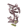
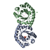
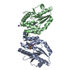
| Deposited unit | 
| ||||||||
|---|---|---|---|---|---|---|---|---|---|
| 1 |
| ||||||||
| Unit cell |
|
-Components
| #1: Protein | Mass: 22662.525 Da / Num. of mol.: 2 Source method: isolated from a genetically manipulated source Source: (gene. exp.) Mycobacterium tuberculosis (strain ATCC 25618 / H37Rv) (bacteria)Strain: ATCC 25618 / H37Rv / Gene: tmk, Rv3247c / Production host: #2: Chemical | 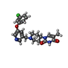  Mass: 426.896 Da / Num. of mol.: 2 / Source method: obtained synthetically / Formula: C22H23ClN4O3 Mass: 426.896 Da / Num. of mol.: 2 / Source method: obtained synthetically / Formula: C22H23ClN4O3#3: Chemical | ChemComp-CL /   Mass: 35.453 Da / Num. of mol.: 4 / Source method: obtained synthetically / Formula: Cl Mass: 35.453 Da / Num. of mol.: 4 / Source method: obtained synthetically / Formula: Cl#4: Water | ChemComp-HOH / |  Mass: 18.015 Da / Num. of mol.: 122 / Source method: isolated from a natural source / Formula: H2O Mass: 18.015 Da / Num. of mol.: 122 / Source method: isolated from a natural source / Formula: H2O |
|---|
-Experimental details
-Experiment
| Experiment | Method: X-RAY DIFFRACTION / Number of used crystals: 1 |
|---|
- Sample preparation
| Crystal | Density Matthews: 2.51 Å3/Da / Density % sol: 51.06 % |
|---|---|
| Crystal grow | Temperature: 293 K / Method: vapor diffusion, sitting drop / Details: 0.1 M HEPES pH 7.5, 4.3 M NaCl |
-Data collection
| Diffraction | Mean temperature: 100 K |
|---|---|
| Diffraction source | Source: SYNCHROTRON / Site: ESRF  / Beamline: ID23-2 / Wavelength: 0.8729 Å / Beamline: ID23-2 / Wavelength: 0.8729 Å |
| Detector | Type: DECTRIS PILATUS 2M-F / Detector: PIXEL / Date: Jun 9, 2016 |
| Radiation | Protocol: SINGLE WAVELENGTH / Monochromatic (M) / Laue (L): M / Scattering type: x-ray |
| Radiation wavelength | Wavelength: 0.8729 Å / Relative weight: 1 |
| Reflection | Resolution: 2.1→50 Å / Num. obs: 25255 / % possible obs: 98 % / Redundancy: 3.65 % / Biso Wilson estimate: 48.9 Å2 / CC1/2: 0.999 / Rrim(I) all: 0.074 / Rsym value: 0.064 / Net I/σ(I): 12.03 |
| Reflection shell | Resolution: 2.1→2.15 Å / Redundancy: 3.45 % / Mean I/σ(I) obs: 1.26 / CC1/2: 0.432 / Rrim(I) all: 1.071 / Rsym value: 0.908 / % possible all: 99.1 |
- Processing
| Software |
| |||||||||||||||||||||||||||||||||||||||||||||||||||||||||||||||||||||||||||
|---|---|---|---|---|---|---|---|---|---|---|---|---|---|---|---|---|---|---|---|---|---|---|---|---|---|---|---|---|---|---|---|---|---|---|---|---|---|---|---|---|---|---|---|---|---|---|---|---|---|---|---|---|---|---|---|---|---|---|---|---|---|---|---|---|---|---|---|---|---|---|---|---|---|---|---|---|
| Refinement | Method to determine structure: MOLECULAR REPLACEMENT Starting model: 4UNR Resolution: 2.1→36.925 Å / SU ML: 0.31 / Cross valid method: FREE R-VALUE / σ(F): 1.92 / Phase error: 24.1 / Stereochemistry target values: ML
| |||||||||||||||||||||||||||||||||||||||||||||||||||||||||||||||||||||||||||
| Solvent computation | Shrinkage radii: 0.9 Å / VDW probe radii: 1.11 Å / Solvent model: FLAT BULK SOLVENT MODEL | |||||||||||||||||||||||||||||||||||||||||||||||||||||||||||||||||||||||||||
| Refinement step | Cycle: LAST / Resolution: 2.1→36.925 Å
| |||||||||||||||||||||||||||||||||||||||||||||||||||||||||||||||||||||||||||
| Refine LS restraints |
| |||||||||||||||||||||||||||||||||||||||||||||||||||||||||||||||||||||||||||
| LS refinement shell |
| |||||||||||||||||||||||||||||||||||||||||||||||||||||||||||||||||||||||||||
| Refinement TLS params. | Method: refined / Refine-ID: X-RAY DIFFRACTION
| |||||||||||||||||||||||||||||||||||||||||||||||||||||||||||||||||||||||||||
| Refinement TLS group |
|
