| Entry | Database: PDB / ID: 5np3
|
|---|
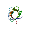
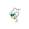
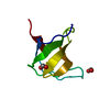
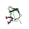
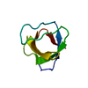
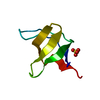
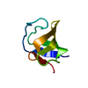
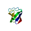
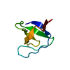
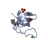
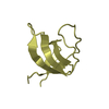
| Title | Abl2 SH3 |
|---|
 Components Components | Abelson tyrosine-protein kinase 2 |
|---|
 Keywords Keywords | TRANSFERASE / signaling / tyrosine phosphorylation / SH3 domain / kinase |
|---|
| Function / homology |  Function and homology information Function and homology information
Role of ABL in ROBO-SLIT signaling / regulation of cell motility / positive regulation of establishment of T cell polarity / negative regulation of Rho protein signal transduction / regulation of endocytosis / exploration behavior / actin monomer binding / RAC3 GTPase cycle / positive regulation of T cell migration / regulation of cell adhesion ...Role of ABL in ROBO-SLIT signaling / regulation of cell motility / positive regulation of establishment of T cell polarity / negative regulation of Rho protein signal transduction / regulation of endocytosis / exploration behavior / actin monomer binding / RAC3 GTPase cycle / positive regulation of T cell migration / regulation of cell adhesion / cellular response to retinoic acid / phosphotyrosine residue binding / peptidyl-tyrosine phosphorylation / RAC1 GTPase cycle / Negative regulation of FLT3 / protein modification process / regulation of autophagy / regulation of actin cytoskeleton organization / non-specific protein-tyrosine kinase / non-membrane spanning protein tyrosine kinase activity / positive regulation of neuron projection development / epidermal growth factor receptor signaling pathway / enzyme activator activity / actin filament binding / manganese ion binding / actin cytoskeleton / positive regulation of cytosolic calcium ion concentration / protein tyrosine kinase activity / cellular response to oxidative stress / phospholipase C-activating G protein-coupled receptor signaling pathway / protein kinase activity / cell adhesion / magnesium ion binding / enzyme binding / signal transduction / ATP binding / plasma membrane / cytosolSimilarity search - Function F-actin binding / F-actin binding / F-actin binding domain (FABD) / Tyrosine-protein kinase ABL, SH2 domain / SH3 Domains / : / SH3 domain / SH2 domain / Src homology 2 (SH2) domain profile. / SH3 type barrels. ...F-actin binding / F-actin binding / F-actin binding domain (FABD) / Tyrosine-protein kinase ABL, SH2 domain / SH3 Domains / : / SH3 domain / SH2 domain / Src homology 2 (SH2) domain profile. / SH3 type barrels. / Src homology 2 domains / SH2 domain / Src homology 3 domains / SH2 domain superfamily / SH3-like domain superfamily / Src homology 3 (SH3) domain profile. / SH3 domain / Tyrosine-protein kinase, catalytic domain / Tyrosine kinase, catalytic domain / Tyrosine protein kinases specific active-site signature. / Tyrosine-protein kinase, active site / Protein tyrosine and serine/threonine kinase / Serine-threonine/tyrosine-protein kinase, catalytic domain / Roll / Protein kinase, ATP binding site / Protein kinases ATP-binding region signature. / Protein kinase domain profile. / Protein kinase domain / Protein kinase-like domain superfamily / Mainly BetaSimilarity search - Domain/homology |
|---|
| Biological species |  Homo sapiens (human) Homo sapiens (human) |
|---|
| Method |  X-RAY DIFFRACTION / SYNCHROTRON / MOLECULAR REPLACEMENT / Resolution: 2 Å X-RAY DIFFRACTION / SYNCHROTRON / MOLECULAR REPLACEMENT / Resolution: 2 Å |
|---|
 Authors Authors | Mero, B. / Radnai, L. / Gogl, G. / Leveles, I. / Buday, L. |
|---|
 Citation Citation | Journal: J.Biol.Chem. / Year: 2019
Title: Structural insights into the tyrosine phosphorylation-mediated inhibition of SH3 domain-ligand interactions.
Authors: Mero, B. / Radnai, L. / Gogl, G. / Toke, O. / Leveles, I. / Koprivanacz, K. / Szeder, B. / Dulk, M. / Kudlik, G. / Vas, V. / Cserkaszky, A. / Sipeki, S. / Nyitray, L. / Vertessy, B.G. / Buday, L. |
|---|
| History | | Deposition | Apr 13, 2017 | Deposition site: PDBE / Processing site: PDBE |
|---|
| Revision 1.0 | May 16, 2018 | Provider: repository / Type: Initial release |
|---|
| Revision 1.1 | Jan 30, 2019 | Group: Data collection / Database references / Category: citation / citation_author
Item: _citation.country / _citation.journal_abbrev ..._citation.country / _citation.journal_abbrev / _citation.journal_id_ASTM / _citation.journal_id_CSD / _citation.journal_id_ISSN / _citation.pdbx_database_id_DOI / _citation.pdbx_database_id_PubMed / _citation.title / _citation.year |
|---|
| Revision 1.2 | Apr 3, 2019 | Group: Data collection / Database references / Category: citation / citation_author / pdbx_database_proc
Item: _citation.journal_abbrev / _citation.journal_volume ..._citation.journal_abbrev / _citation.journal_volume / _citation.page_first / _citation.page_last / _citation_author.identifier_ORCID |
|---|
| Revision 1.3 | Jan 17, 2024 | Group: Data collection / Database references / Refinement description
Category: chem_comp_atom / chem_comp_bond ...chem_comp_atom / chem_comp_bond / database_2 / pdbx_initial_refinement_model
Item: _database_2.pdbx_DOI / _database_2.pdbx_database_accession |
|---|
|
|---|
 Movie
Movie Controller


 Open data
 Basic information
 Structure visualization
 Downloads & links
 Other downloads


 PDBj
PDBj


 Assembly


 Mass: 18.015 Da / Num. of mol.: 37 / Source method: isolated from a natural source / Formula: H2O
 Sample preparation
 / Beamline: P13 (MX1) / Wavelength: 0.9763 Å
 Processing