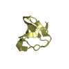
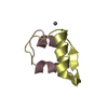
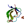
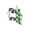
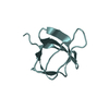
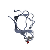
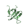
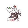
Entry Database : PDB / ID : 5np2Title Abl1 SH3 pTyr89/134 Tyrosine-protein kinase ABL1 Keywords / / / / Function / homology Function Domain/homology Component
/ / / / / / / / / / / / / / / / / / / / / / / / / / / / / / / / / / / / / / / / / / / / / / / / / / / / / / / / / / / / / / / / / / / / / / / / / / / / / / / / / / / / / / / / / / / / / / / / / / / / / / / / / / / / / / / / / / / / / / / / / / / / / / / / / / / / / / / / / / / / / / / / / / Biological species Homo sapiens (human)Method / / / Resolution : 1.6 Å Authors Mero, B. / Radnai, L. / Gogl, G. / Leveles, I. / Buday, L. Journal : J.Biol.Chem. / Year : 2019Title : Structural insights into the tyrosine phosphorylation-mediated inhibition of SH3 domain-ligand interactions.Authors : Mero, B. / Radnai, L. / Gogl, G. / Toke, O. / Leveles, I. / Koprivanacz, K. / Szeder, B. / Dulk, M. / Kudlik, G. / Vas, V. / Cserkaszky, A. / Sipeki, S. / Nyitray, L. / Vertessy, B.G. / Buday, L. History Deposition Apr 13, 2017 Deposition site / Processing site Revision 1.0 May 16, 2018 Provider / Type Revision 1.1 Jan 30, 2019 Group / Database references / Category / citation_authorItem _citation.country / _citation.journal_abbrev ... _citation.country / _citation.journal_abbrev / _citation.journal_id_ASTM / _citation.journal_id_CSD / _citation.journal_id_ISSN / _citation.pdbx_database_id_DOI / _citation.pdbx_database_id_PubMed / _citation.title / _citation.year Revision 1.2 Apr 3, 2019 Group / Database references / Category / citation_author / pdbx_database_procItem _citation.journal_abbrev / _citation.journal_volume ... _citation.journal_abbrev / _citation.journal_volume / _citation.page_first / _citation.page_last / _citation_author.identifier_ORCID Revision 1.3 Jan 17, 2024 Group / Database references / Refinement descriptionCategory chem_comp_atom / chem_comp_bond ... chem_comp_atom / chem_comp_bond / database_2 / pdbx_initial_refinement_model Item / _database_2.pdbx_database_accessionRevision 1.4 Oct 23, 2024 Group / Category / pdbx_modification_feature
Show all Show less
 Movie
Movie Controller


 Open data
 Basic information
 Components
 Keywords
 Function and homology information
 Homo sapiens (human)
 X-RAY DIFFRACTION /
  Authors
 Citation
 Structure visualization
 Downloads & links
 Other downloads


 PDBj
PDBj


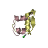
 Assembly


 Mass: 18.015 Da / Num. of mol.: 73 / Source method: isolated from a natural source / Formula: H2O
 Sample preparation
 / Beamline: P13 (MX1) / Wavelength: 0.9763 Å
 Processing