 Movie
Movie Controller

[English] 日本語
 Yorodumi
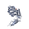
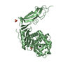
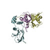
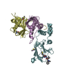
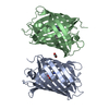
- PDB-5nm9: Crystal structure of the placozoa Trichoplax adhaerens Smad4-MH1 ... -
+ Open data

- Basic information
| Entry | Database: PDB / ID: 5nm9 | ||||||
|---|---|---|---|---|---|---|---|
| Title | Crystal structure of the placozoa Trichoplax adhaerens Smad4-MH1 bound to the GGCGC site. | ||||||
 Components Components |
| ||||||
 Keywords Keywords | TRANSCRIPTION / Smads / transcription factor / DNA complex | ||||||
| Function / homology |  Function and homology information Function and homology informationheteromeric SMAD protein complex / SMAD protein signal transduction / I-SMAD binding / BMP signaling pathway / anatomical structure morphogenesis / DNA-binding transcription factor activity, RNA polymerase II-specific / cell differentiation / RNA polymerase II cis-regulatory region sequence-specific DNA binding / regulation of transcription by RNA polymerase II / metal ion binding / cytoplasm Similarity search - Function | ||||||
| Biological species |   Homo sapiens (human) Homo sapiens (human) | ||||||
| Method |  X-RAY DIFFRACTION / SYNCHROTRON / MOLECULAR REPLACEMENT / Resolution: 2.43 Å X-RAY DIFFRACTION / SYNCHROTRON / MOLECULAR REPLACEMENT / Resolution: 2.43 Å | ||||||
 Authors Authors | Kaczmarska, Z. / Freier, R. / Marquez, J.A. / Macias, M.J. | ||||||
 Citation Citation | Journal: Nat Commun / Year: 2017 Title: Structural basis for genome wide recognition of 5-bp GC motifs by SMAD transcription factors. Authors: Martin-Malpartida, P. / Batet, M. / Kaczmarska, Z. / Freier, R. / Gomes, T. / Aragon, E. / Zou, Y. / Wang, Q. / Xi, Q. / Ruiz, L. / Vea, A. / Marquez, J.A. / Massague, J. / Macias, M.J. | ||||||
| History |
|
- Structure visualization
| Structure viewer | Molecule: MolmilJmol/JSmol |
|---|
- Downloads & links
-Download
| PDBx/mmCIF format | 5nm9.cif.gz | 152.8 KB | Display | PDBx/mmCIF format |
|---|---|---|---|---|
| PDB format | pdb5nm9.ent.gz | 118.1 KB | Display | PDB format |
| PDBx/mmJSON format | 5nm9.json.gz | Tree view | PDBx/mmJSON format | |
| Others |  Other downloads Other downloads |
-Validation report
| Arichive directory | https://data.pdbj.org/pub/pdb/validation_reports/nm/5nm9ftp://data.pdbj.org/pub/pdb/validation_reports/nm/5nm9 | HTTPS FTP |
|---|
-Related structure data
| Related structure data | 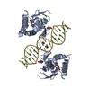 5meySC 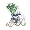 5mezC  5mf0C 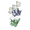 5od6C  5odgC C: citing same article ( S: Starting model for refinement |
|---|---|
| Similar structure data |


-Links
 PDBj
PDBj


- Assembly
| Deposited unit | 
| ||||||||
|---|---|---|---|---|---|---|---|---|---|
| 1 | 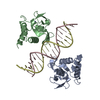
| ||||||||
| 2 | 
| ||||||||
| Unit cell |
|
-Components
| #1: Protein | Mass: 15985.443 Da / Num. of mol.: 2 / Fragment: MH1 domain, UNP residues 33-174 Source method: isolated from a genetically manipulated source Source: (gene. exp.)  #2: DNA chain | Mass: 5518.555 Da / Num. of mol.: 2 / Source method: obtained synthetically / Source: (synth.) Homo sapiens (human)#3: Chemical |   Mass: 65.409 Da / Num. of mol.: 2 / Source method: obtained synthetically / Formula: Zn Mass: 65.409 Da / Num. of mol.: 2 / Source method: obtained synthetically / Formula: Zn#4: Water | ChemComp-HOH / |  Mass: 18.015 Da / Num. of mol.: 19 / Source method: isolated from a natural source / Formula: H2O Mass: 18.015 Da / Num. of mol.: 19 / Source method: isolated from a natural source / Formula: H2O |
|---|
-Experimental details
-Experiment
| Experiment | Method: X-RAY DIFFRACTION / Number of used crystals: 1 |
|---|
- Sample preparation
| Crystal | Density Matthews: 2.41 Å3/Da / Density % sol: 48.99 % |
|---|---|
| Crystal grow | Temperature: 277 K / Method: vapor diffusion, sitting drop / Details: 4.8% PEG 4000, 0.1 M sodium acetate pH 4.6 |
-Data collection
| Diffraction | Mean temperature: 100 K |
|---|---|
| Diffraction source | Source: SYNCHROTRON / Site: ESRF  / Beamline: ID23-1 / Wavelength: 0.97625 Å / Beamline: ID23-1 / Wavelength: 0.97625 Å |
| Detector | Type: ADSC QUANTUM 315r / Detector: CCD / Date: Nov 27, 2016 |
| Radiation | Protocol: SINGLE WAVELENGTH / Monochromatic (M) / Laue (L): M / Scattering type: x-ray |
| Radiation wavelength | Wavelength: 0.97625 Å / Relative weight: 1 |
| Reflection | Biso Wilson estimate: 77.62 Å2 |
| Reflection shell | Resolution: 2.43→2.44 Å / Redundancy: 4.8 % / Mean I/σ(I) obs: 0.8 / Num. unique obs: 204 / % possible all: 100 |
- Processing
| Software |
| |||||||||||||||||||||||||||||||||||||||||||||||||||||||||||||||||||||||||||||||||||||||||||||||||||||||||||||||||||||||||||||
|---|---|---|---|---|---|---|---|---|---|---|---|---|---|---|---|---|---|---|---|---|---|---|---|---|---|---|---|---|---|---|---|---|---|---|---|---|---|---|---|---|---|---|---|---|---|---|---|---|---|---|---|---|---|---|---|---|---|---|---|---|---|---|---|---|---|---|---|---|---|---|---|---|---|---|---|---|---|---|---|---|---|---|---|---|---|---|---|---|---|---|---|---|---|---|---|---|---|---|---|---|---|---|---|---|---|---|---|---|---|---|---|---|---|---|---|---|---|---|---|---|---|---|---|---|---|---|
| Refinement | Method to determine structure: MOLECULAR REPLACEMENT Starting model: 5MEY Resolution: 2.43→29.46 Å / Cor.coef. Fo:Fc: 0.948 / Cor.coef. Fo:Fc free: 0.914 / Rfactor Rfree error: 0 / SU R Cruickshank DPI: 0.369 / Cross valid method: THROUGHOUT / σ(F): 0 / SU R Blow DPI: 0.39 / SU Rfree Blow DPI: 0.248 / SU Rfree Cruickshank DPI: 0.246
| |||||||||||||||||||||||||||||||||||||||||||||||||||||||||||||||||||||||||||||||||||||||||||||||||||||||||||||||||||||||||||||
| Displacement parameters | Biso mean: 82.04 Å2
| |||||||||||||||||||||||||||||||||||||||||||||||||||||||||||||||||||||||||||||||||||||||||||||||||||||||||||||||||||||||||||||
| Refine analyze | Luzzati coordinate error obs: 0.4 Å | |||||||||||||||||||||||||||||||||||||||||||||||||||||||||||||||||||||||||||||||||||||||||||||||||||||||||||||||||||||||||||||
| Refinement step | Cycle: 1 / Resolution: 2.43→29.46 Å
| |||||||||||||||||||||||||||||||||||||||||||||||||||||||||||||||||||||||||||||||||||||||||||||||||||||||||||||||||||||||||||||
| Refine LS restraints |
| |||||||||||||||||||||||||||||||||||||||||||||||||||||||||||||||||||||||||||||||||||||||||||||||||||||||||||||||||||||||||||||
| LS refinement shell | Resolution: 2.43→2.6 Å / Rfactor Rfree error: 0 / Total num. of bins used: 8
| |||||||||||||||||||||||||||||||||||||||||||||||||||||||||||||||||||||||||||||||||||||||||||||||||||||||||||||||||||||||||||||
| Refinement TLS params. | Method: refined / Refine-ID: X-RAY DIFFRACTION
| |||||||||||||||||||||||||||||||||||||||||||||||||||||||||||||||||||||||||||||||||||||||||||||||||||||||||||||||||||||||||||||
| Refinement TLS group |
|
