 Movie
Movie Controller


+ Open data

- Basic information
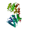
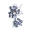
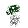
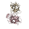
| Entry | Database: PDB / ID: 5n7z | |||||||||
|---|---|---|---|---|---|---|---|---|---|---|
| Title | glycosyltransferase in LPS biosynthesis | |||||||||
 Components Components | Lipopolysaccharide 1,6-galactosyltransferase | |||||||||
 Keywords Keywords | TRANSFERASE / glycosyltransferase LPS biosynthesis | |||||||||
| Function / homology | Glycosyltransferase Family 4 / Glycosyltransferase subfamily 4-like, N-terminal domain / Glycosyl transferase, family 1 / lipopolysaccharide core region biosynthetic process / Glycosyl transferases group 1 / Transferases; Glycosyltransferases; Hexosyltransferases / glycosyltransferase activity / Lipopolysaccharide 1,6-galactosyltransferase Function and homology information Function and homology information | |||||||||
| Biological species |  Salmonella typhimurium (bacteria) Salmonella typhimurium (bacteria) | |||||||||
| Method |  X-RAY DIFFRACTION / SYNCHROTRON / SAD / Resolution: 1.81 Å X-RAY DIFFRACTION / SYNCHROTRON / SAD / Resolution: 1.81 Å | |||||||||
 Authors Authors | Ashworth, G.J. / Zhang, Z. | |||||||||
 Citation Citation | Journal: To Be Published Title: glycosyltransferase in LPS biosynthesis Authors: Ashworth, G.J. / Zhang, Z. | |||||||||
| History |
|
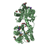
- Structure visualization
| Structure viewer | Molecule: MolmilJmol/JSmol |
|---|
- Downloads & links
-Download
| PDBx/mmCIF format | 5n7z.cif.gz | 84.5 KB | Display | PDBx/mmCIF format |
|---|---|---|---|---|
| PDB format | pdb5n7z.ent.gz | 63.5 KB | Display | PDB format |
| PDBx/mmJSON format | 5n7z.json.gz | Tree view | PDBx/mmJSON format | |
| Others |  Other downloads Other downloads |
-Validation report
| Arichive directory | https://data.pdbj.org/pub/pdb/validation_reports/n7/5n7zftp://data.pdbj.org/pub/pdb/validation_reports/n7/5n7z | HTTPS FTP |
|---|
-Related structure data
| Similar structure data |
|---|


-Links
 PDBj
PDBj
- Assembly
| Deposited unit | 
| ||||||||
|---|---|---|---|---|---|---|---|---|---|
| 1 |
| ||||||||
| Unit cell |
| ||||||||
| Components on special symmetry positions |
|
-Components
| #1: Protein | Mass: 40943.172 Da / Num. of mol.: 1 Source method: isolated from a genetically manipulated source Source: (gene. exp.) Salmonella typhimurium (strain LT2 / SGSC1412 / ATCC 700720) (bacteria)Strain: LT2 / SGSC1412 / ATCC 700720 / Gene: rfaB, waaB, STM3719 / Production host: References: UniProt: Q06994, Transferases; Glycosyltransferases; Hexosyltransferases |
|---|---|
| #2: Water | ChemComp-HOH /  Mass: 18.015 Da / Num. of mol.: 92 / Source method: isolated from a natural source / Formula: H2O Mass: 18.015 Da / Num. of mol.: 92 / Source method: isolated from a natural source / Formula: H2O |
-Experimental details
-Experiment
| Experiment | Method: X-RAY DIFFRACTION / Number of used crystals: 1 |
|---|
- Sample preparation
| Crystal | Density Matthews: 2.94 Å3/Da / Density % sol: 58.1 % |
|---|---|
| Crystal grow | Temperature: 298.15 K / Method: evaporation Details: 1% trypton; 0.001M sodium azide; 0.05M HEPES pH7.0 and 20% w/v PEG 3350 |
-Data collection
| Diffraction | Mean temperature: 77 K |
|---|---|
| Diffraction source | Source: SYNCHROTRON / Site: Diamond  / Beamline: I04 / Wavelength: 1.8233 Å / Beamline: I04 / Wavelength: 1.8233 Å |
| Detector | Type: ADSC QUANTUM 315 / Detector: CCD / Date: May 18, 2015 |
| Radiation | Protocol: SINGLE WAVELENGTH / Monochromatic (M) / Laue (L): M / Scattering type: x-ray |
| Radiation wavelength | Wavelength: 1.8233 Å / Relative weight: 1 |
| Reflection | Resolution: 1.81→67.64 Å / Num. obs: 44340 / % possible obs: 98.5 % / Redundancy: 18.2 % / Net I/σ(I): 3.1 |
- Processing
| Software |
| |||||||||||||||||||||||||||||||||||||||||||||||||||||||||||||||||||||||||||||||||||||||||||||||||||||||||||||||||||||||
|---|---|---|---|---|---|---|---|---|---|---|---|---|---|---|---|---|---|---|---|---|---|---|---|---|---|---|---|---|---|---|---|---|---|---|---|---|---|---|---|---|---|---|---|---|---|---|---|---|---|---|---|---|---|---|---|---|---|---|---|---|---|---|---|---|---|---|---|---|---|---|---|---|---|---|---|---|---|---|---|---|---|---|---|---|---|---|---|---|---|---|---|---|---|---|---|---|---|---|---|---|---|---|---|---|---|---|---|---|---|---|---|---|---|---|---|---|---|---|---|---|
| Refinement | Method to determine structure: SAD / Resolution: 1.81→67.446 Å / SU ML: 0.23 / Cross valid method: NONE / σ(F): 2.03 / Phase error: 24.94
| |||||||||||||||||||||||||||||||||||||||||||||||||||||||||||||||||||||||||||||||||||||||||||||||||||||||||||||||||||||||
| Solvent computation | Shrinkage radii: 0.9 Å / VDW probe radii: 1.11 Å | |||||||||||||||||||||||||||||||||||||||||||||||||||||||||||||||||||||||||||||||||||||||||||||||||||||||||||||||||||||||
| Refinement step | Cycle: LAST / Resolution: 1.81→67.446 Å
| |||||||||||||||||||||||||||||||||||||||||||||||||||||||||||||||||||||||||||||||||||||||||||||||||||||||||||||||||||||||
| Refine LS restraints |
| |||||||||||||||||||||||||||||||||||||||||||||||||||||||||||||||||||||||||||||||||||||||||||||||||||||||||||||||||||||||
| LS refinement shell |
|
