 Movie
Movie Controller

[English] 日本語
 Yorodumi
- PDB-6y6i: lipopolysaccharide outer core galactosyltransferase WaaB and UDP ... -
+ Open data

- Basic information
| Entry | Database: PDB / ID: 6y6i | ||||||
|---|---|---|---|---|---|---|---|
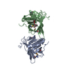
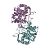
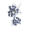
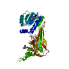
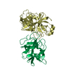
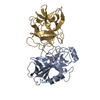
| Title | lipopolysaccharide outer core galactosyltransferase WaaB and UDP complex | ||||||
 Components Components | Lipopolysaccharide 1,6-galactosyltransferase | ||||||
 Keywords Keywords | BIOSYNTHETIC PROTEIN / WaaB / lipopolysaccharide / galactosyltransferase / UDP / lipopolysaccharide outer core | ||||||
| Function / homology | Glycosyltransferase Family 4 / Glycosyltransferase subfamily 4-like, N-terminal domain / Glycosyl transferase, family 1 / lipopolysaccharide core region biosynthetic process / Glycosyl transferases group 1 / Transferases; Glycosyltransferases; Hexosyltransferases / glycosyltransferase activity / URIDINE-5'-DIPHOSPHATE / Lipopolysaccharide 1,6-galactosyltransferase Function and homology information Function and homology information | ||||||
| Biological species |  Salmonella typhimurium (bacteria) Salmonella typhimurium (bacteria) | ||||||
| Method |  X-RAY DIFFRACTION / SYNCHROTRON / MOLECULAR REPLACEMENT / Resolution: 1.92 Å X-RAY DIFFRACTION / SYNCHROTRON / MOLECULAR REPLACEMENT / Resolution: 1.92 Å | ||||||
 Authors Authors | Zhang, Z.Y. / Ashworth, G. / Wang, Z.S. / Zhu, X.F. / Dong, C.J. | ||||||
| Funding support |  United Kingdom, 1items United Kingdom, 1items
| ||||||
 Citation Citation | Journal: To Be Published Title: Structural insights into the lipopolysaccharide outer core galactosyltransferase WaaB Authors: Zhang, Z.Y. / Ashworth, G. / Wang, Z.S. / Zhu, X.F. / Dong, C.J. | ||||||
| History |
|
- Structure visualization
| Structure viewer | Molecule: MolmilJmol/JSmol |
|---|
- Downloads & links
-Download
| PDBx/mmCIF format | 6y6i.cif.gz | 174.9 KB | Display | PDBx/mmCIF format |
|---|---|---|---|---|
| PDB format | pdb6y6i.ent.gz | 136.2 KB | Display | PDB format |
| PDBx/mmJSON format | 6y6i.json.gz | Tree view | PDBx/mmJSON format | |
| Others |  Other downloads Other downloads |
-Validation report
| Arichive directory | https://data.pdbj.org/pub/pdb/validation_reports/y6/6y6iftp://data.pdbj.org/pub/pdb/validation_reports/y6/6y6i | HTTPS FTP |
|---|
-Related structure data
| Related structure data | 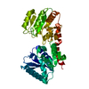 6y6gSC S: Starting model for refinement C: citing same article ( |
|---|---|
| Similar structure data |


-Links
 PDBj
PDBj


- Assembly
| Deposited unit | 
| ||||||||
|---|---|---|---|---|---|---|---|---|---|
| 1 |
| ||||||||
| Unit cell |
| ||||||||
| Components on special symmetry positions |
|
-Components
| #1: Protein | Mass: 41071.301 Da / Num. of mol.: 1 Source method: isolated from a genetically manipulated source Source: (gene. exp.) Salmonella typhimurium (strain LT2 / SGSC1412 / ATCC 700720) (bacteria)Gene: rfaB, waaB, STM3719 / Production host: References: UniProt: Q06994, Transferases; Glycosyltransferases; Hexosyltransferases |
|---|---|
| #2: Chemical | ChemComp-UDP /   Type: RNA linking / Mass: 404.161 Da / Num. of mol.: 1 / Source method: obtained synthetically / Formula: C9H14N2O12P2 / Feature type: SUBJECT OF INVESTIGATION / Comment: UDP*YM Type: RNA linking / Mass: 404.161 Da / Num. of mol.: 1 / Source method: obtained synthetically / Formula: C9H14N2O12P2 / Feature type: SUBJECT OF INVESTIGATION / Comment: UDP*YM |
| #3: Water | ChemComp-HOH /  Mass: 18.015 Da / Num. of mol.: 326 / Source method: isolated from a natural source / Formula: H2O Mass: 18.015 Da / Num. of mol.: 326 / Source method: isolated from a natural source / Formula: H2O |
| Has ligand of interest | Y |
-Experimental details
-Experiment
| Experiment | Method: X-RAY DIFFRACTION / Number of used crystals: 1 |
|---|
- Sample preparation
| Crystal | Density Matthews: 2.98 Å3/Da / Density % sol: 58.67 % |
|---|---|
| Crystal grow | Temperature: 293 K / Method: vapor diffusion, sitting drop / pH: 7 Details: 1% tryptone, 1mM sodium azide, 0.05M HEPES pH7.0 and 20% w/v PEG 3350 |
-Data collection
| Diffraction | Mean temperature: 81 K / Serial crystal experiment: N |
|---|---|
| Diffraction source | Source: SYNCHROTRON / Site: Diamond / Beamline: I03 / Wavelength: 0.979 Å |
| Detector | Type: ADSC QUANTUM 4 / Detector: CCD / Date: Feb 9, 2015 |
| Radiation | Protocol: SINGLE WAVELENGTH / Monochromatic (M) / Laue (L): M / Scattering type: x-ray |
| Radiation wavelength | Wavelength: 0.979 Å / Relative weight: 1 |
| Reflection | Resolution: 1.92→73.85 Å / Num. obs: 38400 / % possible obs: 99.9 % / Redundancy: 23.4 % / CC1/2: 1 / Rmerge(I) obs: 0.16 / Net I/σ(I): 16.1 |
| Reflection shell | Resolution: 1.92→1.989 Å / Redundancy: 11.8 % / Rmerge(I) obs: 1.33 / Num. unique obs: 3752 / CC1/2: 0.5 / % possible all: 99.5 |
- Processing
| Software |
| |||||||||||||||||||||||||||||||||||||||||||||||||||||||||||||||||
|---|---|---|---|---|---|---|---|---|---|---|---|---|---|---|---|---|---|---|---|---|---|---|---|---|---|---|---|---|---|---|---|---|---|---|---|---|---|---|---|---|---|---|---|---|---|---|---|---|---|---|---|---|---|---|---|---|---|---|---|---|---|---|---|---|---|---|
| Refinement | Method to determine structure: MOLECULAR REPLACEMENT Starting model: 6Y6G Resolution: 1.92→73.85 Å / Cor.coef. Fo:Fc: 0.965 / Cor.coef. Fo:Fc free: 0.946 / SU B: 8.067 / SU ML: 0.1 / Cross valid method: THROUGHOUT / σ(F): 0 / ESU R: 0.207 / ESU R Free: 0.124 Details: HYDROGENS HAVE BEEN ADDED IN THE RIDING POSITIONS U VALUES : WITH TLS ADDED
| |||||||||||||||||||||||||||||||||||||||||||||||||||||||||||||||||
| Solvent computation | Ion probe radii: 0.8 Å / Shrinkage radii: 0.8 Å / VDW probe radii: 1.2 Å | |||||||||||||||||||||||||||||||||||||||||||||||||||||||||||||||||
| Displacement parameters | Biso max: 120.3 Å2 / Biso mean: 33.397 Å2 / Biso min: 17.96 Å2
| |||||||||||||||||||||||||||||||||||||||||||||||||||||||||||||||||
| Refinement step | Cycle: final / Resolution: 1.92→73.85 Å
| |||||||||||||||||||||||||||||||||||||||||||||||||||||||||||||||||
| Refine LS restraints |
| |||||||||||||||||||||||||||||||||||||||||||||||||||||||||||||||||
| LS refinement shell | Resolution: 1.92→1.97 Å / Rfactor Rfree error: 0 / Total num. of bins used: 20
| |||||||||||||||||||||||||||||||||||||||||||||||||||||||||||||||||
| Refinement TLS params. | Method: refined / Origin x: -26.5 Å / Origin y: 2.321 Å / Origin z: -19.749 Å
|
