caveola neck / negative regulation of protein processing involved in protein targeting to mitochondrion / Wnt signalosome assembly / beta-catenin destruction complex binding / regulation of branching morphogenesis of a nerve / regulation of kidney size / regulation of cell projection organization / tangential migration from the subventricular zone to the olfactory bulb / protein localization to endoplasmic reticulum exit site / GTP-dependent protein kinase activity ...caveola neck / negative regulation of protein processing involved in protein targeting to mitochondrion / Wnt signalosome assembly / beta-catenin destruction complex binding / regulation of branching morphogenesis of a nerve / regulation of kidney size / regulation of cell projection organization / tangential migration from the subventricular zone to the olfactory bulb / protein localization to endoplasmic reticulum exit site / GTP-dependent protein kinase activity / regulation of SNARE complex assembly / regulation of neuroblast proliferation / regulation of ER to Golgi vesicle-mediated transport / peroxidase inhibitor activity / negative regulation of late endosome to lysosome transport / regulation of mitochondrial depolarization / negative regulation of protein targeting to mitochondrion / positive regulation of dopamine receptor signaling pathway / amphisome / regulation of synaptic vesicle transport / regulation of CAMKK-AMPK signaling cascade / regulation of lysosomal lumen pH / co-receptor binding / negative regulation of GTPase activity / regulation of dopamine receptor signaling pathway / regulation of neuron maturation / positive regulation of microglial cell activation / regulation of retrograde transport, endosome to Golgi / positive regulation of synaptic vesicle endocytosis / cytoplasmic side of mitochondrial outer membrane / negative regulation of excitatory postsynaptic potential / negative regulation of autophagosome assembly / JUN kinase kinase kinase activity / olfactory bulb development / neuron projection arborization / multivesicular body, internal vesicle / striatum development / mitochondrion localization / regulation of dendritic spine morphogenesis / protein localization to mitochondrion / cellular response to dopamine / positive regulation of mitochondrial outer membrane permeabilization involved in apoptotic signaling pathway / endoplasmic reticulum organization / positive regulation of protein autoubiquitination / Wnt signalosome / negative regulation of protein processing / positive regulation of programmed cell death / GTP metabolic process / regulation of canonical Wnt signaling pathway / syntaxin-1 binding / regulation of reactive oxygen species metabolic process / lysosome organization / Golgi-associated vesicle / clathrin binding / protein kinase A binding / PTK6 promotes HIF1A stabilization / negative regulation of macroautophagy / regulation of locomotion / regulation of epidermal cell division / neuromuscular junction development / protein kinase C inhibitor activity / regulation of cAMP/PKA signal transduction / positive regulation of epidermal cell differentiation / keratinocyte development / regulation of mitochondrial fission / keratinization / Golgi organization / regulation of synaptic vesicle exocytosis / microvillus / exploration behavior / intracellular distribution of mitochondria / regulation of cell-cell adhesion / endoplasmic reticulum exit site / autolysosome / locomotory exploration behavior / cAMP/PKA signal transduction / Regulation of localization of FOXO transcription factors / keratinocyte proliferation / negative regulation of Notch signaling pathway / Activation of BAD and translocation to mitochondria / phosphoserine residue binding / MAP kinase kinase kinase activity / regulation of synaptic vesicle endocytosis / negative regulation of keratinocyte proliferation / canonical Wnt signaling pathway / establishment of skin barrier / negative regulation of protein localization to plasma membrane / Chk1/Chk2(Cds1) mediated inactivation of Cyclin B:Cdk1 complex / SARS-CoV-2 targets host intracellular signalling and regulatory pathways / regulation of synaptic transmission, glutamatergic / negative regulation of endoplasmic reticulum stress-induced intrinsic apoptotic signaling pathway / negative regulation of protein kinase activity / negative regulation of stem cell proliferation / presynaptic cytosol / Rho protein signal transduction / SARS-CoV-1 targets host intracellular signalling and regulatory pathways / RHO GTPases activate PKNs / phagocytic vesicle / neuron projection morphogenesis / positive regulation of protein localization Similarity search - Function
: / : / : / LRRK2 ARM repeat / LRRK2 ANK repeat / LRRK2 beta propeller / : / C-terminal of Roc, COR-B domain / C-terminal of Roc (COR) domain / C-terminal of Roc, COR-A domain ...: / : / : / LRRK2 ARM repeat / LRRK2 ANK repeat / LRRK2 beta propeller / : / C-terminal of Roc, COR-B domain / C-terminal of Roc (COR) domain / C-terminal of Roc, COR-A domain / Ras of Complex, Roc, domain of DAPkinase / Roc domain profile. / Roc domain / 14-3-3 domain / Delta-Endotoxin; domain 1 / Leucine-rich repeats, bacterial type / 14-3-3 protein sigma / 14-3-3 proteins signature 2. / 14-3-3 protein, conserved site / 14-3-3 proteins signature 1. / 14-3-3 protein / 14-3-3 homologues / 14-3-3 domain / 14-3-3 domain superfamily / 14-3-3 protein / Leucine rich repeat / Leucine-rich repeat, typical subtype / Leucine-rich repeats, typical (most populated) subfamily / Leucine-rich repeat profile. / Leucine-rich repeat / Rab subfamily of small GTPases / Leucine-rich repeat domain superfamily / Ankyrin repeat-containing domain superfamily / Armadillo-like helical / Small GTP-binding protein domain / Armadillo-type fold / Serine/threonine-protein kinase, active site / Serine/Threonine protein kinases active-site signature. / Protein kinase domain / WD40-repeat-containing domain superfamily / Serine/Threonine protein kinases, catalytic domain / WD40/YVTN repeat-like-containing domain superfamily / Protein kinase, ATP binding site / Protein kinases ATP-binding region signature. / Protein kinase domain profile. / Protein kinase domain / Protein kinase-like domain superfamily / Up-down Bundle / P-loop containing nucleoside triphosphate hydrolase / Mainly Alpha Similarity search - Domain/homology
In the structure databanks used in Yorodumi, some data are registered as the other names, "COVID-19 virus" and "2019-nCoV". Here are the details of the virus and the list of structure data.
Jan 31, 2019. EMDB accession codes are about to change! (news from PDBe EMDB page)
EMDB accession codes are about to change! (news from PDBe EMDB page)
The allocation of 4 digits for EMDB accession codes will soon come to an end. Whilst these codes will remain in use, new EMDB accession codes will include an additional digit and will expand incrementally as the available range of codes is exhausted. The current 4-digit format prefixed with “EMD-” (i.e. EMD-XXXX) will advance to a 5-digit format (i.e. EMD-XXXXX), and so on. It is currently estimated that the 4-digit codes will be depleted around Spring 2019, at which point the 5-digit format will come into force.
The EM Navigator/Yorodumi systems omit the EMD- prefix.
Related info.:Q: What is EMD? / ID/Accession-code notation in Yorodumi/EM Navigator
Yorodumi is a browser for structure data from EMDB, PDB, SASBDB, etc.
This page is also the successor to EM Navigator detail page, and also detail information page/front-end page for Omokage search.
The word "yorodu" (or yorozu) is an old Japanese word meaning "ten thousand". "mi" (miru) is to see.
Related info.:EMDB / PDB / SASBDB / Comparison of 3 databanks / Yorodumi Search / Aug 31, 2016. New EM Navigator & Yorodumi / Yorodumi Papers / Jmol/JSmol / Function and homology information / Changes in new EM Navigator and Yorodumi
 Movie
Movie Controller

 Yorodumi
 Open data
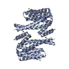
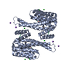
 Basic information
 Components
 Keywords
 Function and homology information
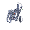
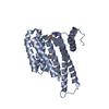
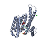
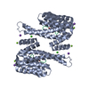
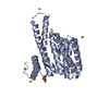
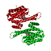
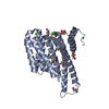
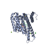
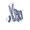
 Homo sapiens (human)
 X-RAY DIFFRACTION /
  Authors
 Citation
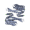
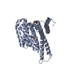
 Structure visualization
 Downloads & links
 Other downloads
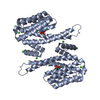


 PDBj
PDBj


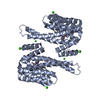
 Assembly
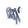


 Mass: 40.078 Da / Num. of mol.: 2 / Source method: obtained synthetically / Formula: Ca
 Mass: 35.453 Da / Num. of mol.: 1 / Source method: obtained synthetically / Formula: Cl
 Mass: 22.990 Da / Num. of mol.: 1 / Source method: obtained synthetically / Formula: Na
 Sample preparation
 / Beamline: X06SA / Wavelength: 0.97862 Å
 Processing