- PDB-5btv: Crystal structure of human 14-3-3 sigma in complex with a Tau-pro... -
+
Open data
ID or keywords:
Loading...
-
Basic information
Entry
Database: PDB / ID: 5btv
Title
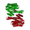
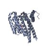
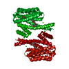
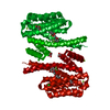
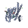
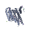
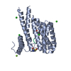
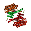
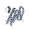
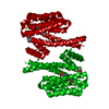
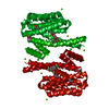
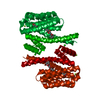
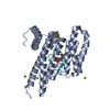
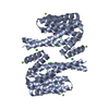
Crystal structure of human 14-3-3 sigma in complex with a Tau-protein peptide surrounding pS324
Components
14-3-3 protein sigma
Microtubule-associated protein tau - peptide pS324
Keywords
STRUCTURAL PROTEIN / Peptide binding protein / 14-3-3 / Tau-protein
Function / homology
Function and homology information
plus-end-directed organelle transport along microtubule / histone-dependent DNA binding / negative regulation of protein localization to mitochondrion / neurofibrillary tangle / microtubule lateral binding / axonal transport / tubulin complex / positive regulation of protein localization to synapse / regulation of epidermal cell division / phosphatidylinositol bisphosphate binding ...plus-end-directed organelle transport along microtubule / histone-dependent DNA binding / negative regulation of protein localization to mitochondrion / neurofibrillary tangle / microtubule lateral binding / axonal transport / tubulin complex / positive regulation of protein localization to synapse / regulation of epidermal cell division / phosphatidylinositol bisphosphate binding / protein kinase C inhibitor activity / generation of neurons / positive regulation of epidermal cell differentiation / keratinocyte development / keratinization / rRNA metabolic process / axonal transport of mitochondrion / regulation of mitochondrial fission / axon development / regulation of microtubule-based movement / intracellular distribution of mitochondria / regulation of chromosome organization / central nervous system neuron development / regulation of cell-cell adhesion / minor groove of adenine-thymine-rich DNA binding / lipoprotein particle binding / microtubule polymerization / negative regulation of mitochondrial membrane potential / establishment of skin barrier / regulation of microtubule polymerization / Regulation of localization of FOXO transcription factors / keratinocyte proliferation / dynactin binding / apolipoprotein binding / protein polymerization / main axon / negative regulation of keratinocyte proliferation / Activation of BAD and translocation to mitochondria / phosphoserine residue binding / Caspase-mediated cleavage of cytoskeletal proteins / cAMP/PKA signal transduction / regulation of microtubule polymerization or depolymerization / negative regulation of protein localization to plasma membrane / negative regulation of mitochondrial fission / axolemma / glial cell projection / SARS-CoV-2 targets host intracellular signalling and regulatory pathways / neurofibrillary tangle assembly / negative regulation of protein kinase activity / negative regulation of stem cell proliferation / positive regulation of axon extension / SARS-CoV-1 targets host intracellular signalling and regulatory pathways / RHO GTPases activate PKNs / Chk1/Chk2(Cds1) mediated inactivation of Cyclin B:Cdk1 complex / regulation of cellular response to heat / positive regulation of microtubule polymerization / positive regulation of protein localization / Activation of AMPK downstream of NMDARs / positive regulation of superoxide anion generation / cellular response to brain-derived neurotrophic factor stimulus / regulation of long-term synaptic depression / supramolecular fiber organization / cytoplasmic microtubule organization / regulation of calcium-mediated signaling / axon cytoplasm / somatodendritic compartment / protein export from nucleus / synapse assembly / phosphatidylinositol binding / nuclear periphery / TP53 Regulates Transcription of Genes Involved in G2 Cell Cycle Arrest / negative regulation of innate immune response / positive regulation of cell adhesion / astrocyte activation / release of cytochrome c from mitochondria / enzyme inhibitor activity / protein phosphatase 2A binding / positive regulation of protein export from nucleus / stem cell proliferation / stress granule assembly / regulation of microtubule cytoskeleton organization / regulation of autophagy / TP53 Regulates Metabolic Genes / Translocation of SLC2A4 (GLUT4) to the plasma membrane / cellular response to reactive oxygen species / protein sequestering activity / microglial cell activation / cellular response to nerve growth factor stimulus / Hsp90 protein binding / protein homooligomerization / SH3 domain binding / PKR-mediated signaling / synapse organization / regulation of synaptic plasticity / response to lead ion / microtubule cytoskeleton organization / intrinsic apoptotic signaling pathway in response to DNA damage / memory / neuron projection development / cytoplasmic ribonucleoprotein granule Similarity search - Function
14-3-3 domain / Delta-Endotoxin; domain 1 / Microtubule-associated protein Tau / Microtubule associated protein, tubulin-binding repeat / Tau and MAP protein, tubulin-binding repeat / Tau and MAP proteins tubulin-binding repeat signature. / Tau and MAP proteins tubulin-binding repeat profile. / : / 14-3-3 protein sigma / 14-3-3 proteins signature 2. ...14-3-3 domain / Delta-Endotoxin; domain 1 / Microtubule-associated protein Tau / Microtubule associated protein, tubulin-binding repeat / Tau and MAP protein, tubulin-binding repeat / Tau and MAP proteins tubulin-binding repeat signature. / Tau and MAP proteins tubulin-binding repeat profile. / : / 14-3-3 protein sigma / 14-3-3 proteins signature 2. / 14-3-3 protein, conserved site / 14-3-3 proteins signature 1. / 14-3-3 protein / 14-3-3 homologues / 14-3-3 domain / 14-3-3 domain superfamily / 14-3-3 protein / Up-down Bundle / Mainly Alpha Similarity search - Domain/homology
In the structure databanks used in Yorodumi, some data are registered as the other names, "COVID-19 virus" and "2019-nCoV". Here are the details of the virus and the list of structure data.
Jan 31, 2019. EMDB accession codes are about to change! (news from PDBe EMDB page)
EMDB accession codes are about to change! (news from PDBe EMDB page)
The allocation of 4 digits for EMDB accession codes will soon come to an end. Whilst these codes will remain in use, new EMDB accession codes will include an additional digit and will expand incrementally as the available range of codes is exhausted. The current 4-digit format prefixed with “EMD-” (i.e. EMD-XXXX) will advance to a 5-digit format (i.e. EMD-XXXXX), and so on. It is currently estimated that the 4-digit codes will be depleted around Spring 2019, at which point the 5-digit format will come into force.
The EM Navigator/Yorodumi systems omit the EMD- prefix.
Related info.:Q: What is EMD? / ID/Accession-code notation in Yorodumi/EM Navigator
Yorodumi is a browser for structure data from EMDB, PDB, SASBDB, etc.
This page is also the successor to EM Navigator detail page, and also detail information page/front-end page for Omokage search.
The word "yorodu" (or yorozu) is an old Japanese word meaning "ten thousand". "mi" (miru) is to see.
Related info.:EMDB / PDB / SASBDB / Comparison of 3 databanks / Yorodumi Search / Aug 31, 2016. New EM Navigator & Yorodumi / Yorodumi Papers / Jmol/JSmol / Function and homology information / Changes in new EM Navigator and Yorodumi
 Movie
Movie Controller

 Yorodumi
 Open data
 Basic information
 Components
 Keywords
 Function and homology information
 Homo sapiens (human)
 X-RAY DIFFRACTION / Resolution: 1.7 Å
  Authors
 Citation
 Structure visualization
 Downloads & links
 Other downloads
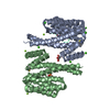


 PDBj
PDBj


 Assembly
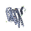


 Mass: 24.305 Da / Num. of mol.: 2 / Source method: obtained synthetically / Formula: Mg
 Mass: 40.078 Da / Num. of mol.: 1 / Source method: obtained synthetically / Formula: Ca
 Mass: 35.453 Da / Num. of mol.: 1 / Source method: obtained synthetically / Formula: Cl
 Sample preparation
 Processing