[English] 日本語
Yorodumi
- PDB-3t0m: Small-molecule inhibitors of 14-3-3 protein-protein interactions ... -

+
Open data


ID or keywords:

Loading...

-
Basic information

Entry
Database: PDB / ID: 3t0m
Title
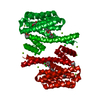
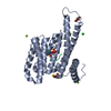
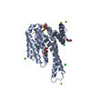
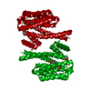
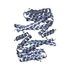
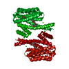
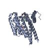
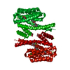
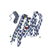
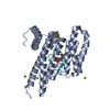
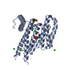
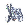
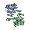
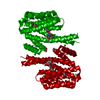
Small-molecule inhibitors of 14-3-3 protein-protein interactions from virtual screening
Components14-3-3 protein sigma
KeywordsPROTEIN BINDING/INHIBITOR / Helical protein / Adapter protein / Protein-protein interaction / PROTEIN BINDING-INHIBITOR complex
Function / homology
Function and homology information


regulation of epidermal cell division / protein kinase C inhibitor activity / positive regulation of epidermal cell differentiation / keratinocyte development / keratinization / Regulation of localization of FOXO transcription factors / keratinocyte proliferation / phosphoserine residue binding / Activation of BAD and translocation to mitochondria / negative regulation of keratinocyte proliferation ...regulation of epidermal cell division / protein kinase C inhibitor activity / positive regulation of epidermal cell differentiation / keratinocyte development / keratinization / Regulation of localization of FOXO transcription factors / keratinocyte proliferation / phosphoserine residue binding / Activation of BAD and translocation to mitochondria / negative regulation of keratinocyte proliferation / establishment of skin barrier / SARS-CoV-2 targets host intracellular signalling and regulatory pathways / Chk1/Chk2(Cds1) mediated inactivation of Cyclin B:Cdk1 complex / protein kinase A signaling / negative regulation of stem cell proliferation / SARS-CoV-1 targets host intracellular signalling and regulatory pathways / RHO GTPases activate PKNs / protein export from nucleus / negative regulation of innate immune response / protein sequestering activity / TP53 Regulates Transcription of Genes Involved in G2 Cell Cycle Arrest / release of cytochrome c from mitochondria / positive regulation of protein export from nucleus / stem cell proliferation / Translocation of SLC2A4 (GLUT4) to the plasma membrane / TP53 Regulates Metabolic Genes / negative regulation of protein kinase activity / negative regulation of cysteine-type endopeptidase activity involved in apoptotic process / intrinsic apoptotic signaling pathway in response to DNA damage / positive regulation of cell growth / regulation of cell cycle / cadherin binding / protein kinase binding / negative regulation of transcription by RNA polymerase II / signal transduction / extracellular space / extracellular exosome / identical protein binding / nucleus / cytosol / cytoplasm
Similarity search - Function
14-3-3 domain / Delta-Endotoxin; domain 1 / 14-3-3 protein sigma / 14-3-3 proteins signature 2. / 14-3-3 protein, conserved site / 14-3-3 proteins signature 1. / 14-3-3 protein / 14-3-3 homologues / 14-3-3 domain / 14-3-3 domain superfamily ...14-3-3 domain / Delta-Endotoxin; domain 1 / 14-3-3 protein sigma / 14-3-3 proteins signature 2. / 14-3-3 protein, conserved site / 14-3-3 proteins signature 1. / 14-3-3 protein / 14-3-3 homologues / 14-3-3 domain / 14-3-3 domain superfamily / 14-3-3 protein / Up-down Bundle / Mainly Alpha
Similarity search - Domain/homology
Chem-2CT / 14-3-3 protein sigma
Similarity search - Component
Biological speciesHomo sapiens (human)
MethodX-RAY DIFFRACTION / SYNCHROTRON / MOLECULAR REPLACEMENT / molecular replacement / Resolution: 1.62 Å
AuthorsThiel, P. / Ottmann, C.
CitationJournal: Chem.Commun.(Camb.) / Year: 2013
Title: Virtual screening and experimental validation reveal novel small-molecule inhibitors of 14-3-3 protein-protein interactions.
Authors: Thiel, P. / Roglin, L. / Meissner, N. / Hennig, S. / Kohlbacher, O. / Ottmann, C.
History
DepositionJul 20, 2011Deposition site: RCSB / Processing site: RCSB
Revision 1.0Dec 26, 2012Provider: repository / Type: Initial release
Revision 1.1Sep 4, 2013Group: Database references
Revision 1.2Feb 28, 2024Group: Data collection / Database references / Derived calculations
Category: chem_comp_atom / chem_comp_bond ...chem_comp_atom / chem_comp_bond / database_2 / pdbx_struct_conn_angle / struct_conn / struct_ref_seq_dif / struct_site
Item: _database_2.pdbx_DOI / _database_2.pdbx_database_accession ..._database_2.pdbx_DOI / _database_2.pdbx_database_accession / _pdbx_struct_conn_angle.ptnr1_auth_comp_id / _pdbx_struct_conn_angle.ptnr1_auth_seq_id / _pdbx_struct_conn_angle.ptnr1_label_alt_id / _pdbx_struct_conn_angle.ptnr1_label_asym_id / _pdbx_struct_conn_angle.ptnr1_label_atom_id / _pdbx_struct_conn_angle.ptnr1_label_comp_id / _pdbx_struct_conn_angle.ptnr1_label_seq_id / _pdbx_struct_conn_angle.ptnr3_auth_comp_id / _pdbx_struct_conn_angle.ptnr3_auth_seq_id / _pdbx_struct_conn_angle.ptnr3_label_alt_id / _pdbx_struct_conn_angle.ptnr3_label_asym_id / _pdbx_struct_conn_angle.ptnr3_label_atom_id / _pdbx_struct_conn_angle.ptnr3_label_comp_id / _pdbx_struct_conn_angle.ptnr3_label_seq_id / _pdbx_struct_conn_angle.value / _struct_conn.pdbx_dist_value / _struct_conn.pdbx_ptnr2_label_alt_id / _struct_conn.ptnr1_auth_comp_id / _struct_conn.ptnr1_auth_seq_id / _struct_conn.ptnr1_label_asym_id / _struct_conn.ptnr1_label_atom_id / _struct_conn.ptnr1_label_comp_id / _struct_conn.ptnr1_label_seq_id / _struct_conn.ptnr2_auth_comp_id / _struct_conn.ptnr2_auth_seq_id / _struct_conn.ptnr2_label_asym_id / _struct_conn.ptnr2_label_atom_id / _struct_conn.ptnr2_label_comp_id / _struct_ref_seq_dif.details / _struct_site.pdbx_auth_asym_id / _struct_site.pdbx_auth_comp_id / _struct_site.pdbx_auth_seq_id

-
Structure visualization

Structure viewerMolecule:
MolmilJmol/JSmol

Downloads & links

-
Assembly

Deposited unit
A: 14-3-3 protein sigma
hetero molecules


Theoretical massNumber of molelcules
Total (without water)27,1819
Polymers26,4861
Non-polymers6958
Water5,999333
1
A: 14-3-3 protein sigma
hetero molecules

A: 14-3-3 protein sigma
hetero molecules


Theoretical massNumber of molelcules
Total (without water)54,36218
Polymers52,9722
Non-polymers1,39116
Water362
TypeNameSymmetry operationNumber
identity operation1_555x,y,z1
crystal symmetry operation4_555x,-y,-z1
Buried area4020 Å2
ΔGint-74 kcal/mol
Surface area22560 Å2
MethodPISA
Unit cell
Length a, b, c (Å)82.090, 112.300, 62.370
Angle α, β, γ (deg.)90.00, 90.00, 90.00
Int Tables number20
Space group name H-MC2221
Components on special symmetry positions
IDModelComponents
11A-503-

HOH

-
Components

-
Protein , 1 types, 1 molecules A

#1: Protein 14-3-3 protein sigma / Epithelial cell marker protein 1 / Stratifin


Mass: 26485.865 Da / Num. of mol.: 1 / Fragment: UNP residues 1-231
Source method: isolated from a genetically manipulated source
Source: (gene. exp.) Homo sapiens (human) / Gene: HME1, SFN / Plasmid: pPROEX HTB / Production host: Escherichia coli (E. coli) / Strain (production host): BL21(DE3) / References: UniProt: P31947

-
Non-polymers , 5 types, 341 molecules

#2: Chemical ChemComp-2CT / (2-{2-[(2,5-dimethoxyphenyl)amino]-2-oxoethoxy}phenyl)phosphonic acid


Mass: 367.290 Da / Num. of mol.: 1 / Source method: obtained synthetically / Formula: C16H18NO7P
#3: Chemical ChemComp-GOL / GLYCEROL / GLYCERIN / PROPANE-1,2,3-TRIOL / Glycerol


Mass: 92.094 Da / Num. of mol.: 2 / Source method: obtained synthetically / Formula: C3H8O3
#4: Chemical ChemComp-CL / CHLORIDE ION / Chloride


Mass: 35.453 Da / Num. of mol.: 2 / Source method: obtained synthetically / Formula: Cl
#5: Chemical ChemComp-MG / MAGNESIUM ION


Mass: 24.305 Da / Num. of mol.: 3 / Source method: obtained synthetically / Formula: Mg
#6: Water ChemComp-HOH / water / Water


Mass: 18.015 Da / Num. of mol.: 333 / Source method: isolated from a natural source / Formula: H2O

-
Experimental details

-
Experiment

ExperimentMethod: X-RAY DIFFRACTION / Number of used crystals: 2

-
Sample preparation

CrystalDensity Matthews: 2.71 Å3/Da / Density % sol: 54.67 %
Crystal growTemperature: 277 K / Method: vapor diffusion, hanging drop / pH: 7.5
Details: 0.095M HEPES Na salt, 0.19M calcium chloride, 5% glycerol, 25.6% PEG 400, pH 7.5, VAPOR DIFFUSION, HANGING DROP, temperature 277K

-
Data collection

Diffraction
IDMean temperature (K)Crystal-ID
11001
21001
Diffraction source
SourceSiteBeamlineTypeIDWavelength (Å)
SYNCHROTRONSLS X10SA10.9778
ROTATING ANODERIGAKU MICROMAX-007 HF21.5418
Detector
TypeIDDetectorDateDetails
DECTRIS PILATUS 6M1PIXELMay 20, 2011
MAR scanner 345 mm plate2IMAGE PLATEApr 14, 2011Multilayer mirrors (VariMax)
Radiation
IDMonochromatorProtocolMonochromatic (M) / Laue (L)Scattering typeWavelength-ID
1Si(111) monochromator (horizontal), dynamically bendable mirror (vertical)SINGLE WAVELENGTHMx-ray1
2SINGLE WAVELENGTHMx-ray1
Radiation wavelength
IDWavelength (Å)Relative weight
10.97781
21.54181
ReflectionResolution: 1.62→45.42 Å / Num. all: 37003 / Num. obs: 36467 / % possible obs: 98.6 % / Observed criterion σ(F): -3 / Observed criterion σ(I): -3 / Redundancy: 6.73 % / Biso Wilson estimate: 24.424 Å2 / Rmerge(I) obs: 0.09 / Net I/σ(I): 13.87
Reflection shell

Diffraction-ID: 1,2

Resolution (Å)Rmerge(I) obsMean I/σ(I) obsNum. measured obsNum. unique allNum. unique obs% possible all
1.62-1.70.3884.04219904759475996.7
1.7-1.80.2865.28219284779477996.2
1.8-20.2388.48467417084708497.8
2-2.30.14314.58548386707670799.9
2.3-2.60.10218.99314583980398099.9
2.6-30.08621.86247293130313099.9
3-40.0725.55252333418341899.8
4-60.06427.52129771801180199.9
6-80.06227.13321445645699.3
8-100.04929.781256166166100
10-120.05428.335257676100
12-45.420.0527.6674511111194.9

-
Phasing

PhasingMethod: molecular replacement
Phasing MRModel details: Phaser MODE: MR_AUTO
Highest resolutionLowest resolution
Rotation2.5 Å45.42 Å
Translation2.5 Å45.42 Å

-
Processing

Software
NameVersionClassificationNB
XSCALEdata scaling
PHASER2.2.1phasing
REFMACrefinement
PDB_EXTRACT3.1data extraction
XDSdata scaling
XDSdata reduction
RefinementMethod to determine structure: MOLECULAR REPLACEMENT / Resolution: 1.62→45.42 Å / Cor.coef. Fo:Fc: 0.966 / Cor.coef. Fo:Fc free: 0.946 / WRfactor Rfree: 0.1983 / WRfactor Rwork: 0.1574 / Occupancy max: 1 / Occupancy min: 0.12 / FOM work R set: 0.8968 / SU B: 1.468 / SU ML: 0.052 / SU R Cruickshank DPI: 0.0807 / SU Rfree: 0.087 / Cross valid method: THROUGHOUT / σ(F): 0 / ESU R Free: 0.087 / Stereochemistry target values: MAXIMUM LIKELIHOOD / Details: U VALUES : REFINED INDIVIDUALLY
RfactorNum. reflection% reflectionSelection details
Rfree0.1978 1824 5 %RANDOM
Rwork0.1552 ---
all0.1573 34642 --
obs0.1573 34642 100 %-
Solvent computationIon probe radii: 0.8 Å / Shrinkage radii: 0.8 Å / VDW probe radii: 1.4 Å / Solvent model: MASK
Displacement parametersBiso max: 66.54 Å2 / Biso mean: 20.171 Å2 / Biso min: 3.33 Å2
Baniso -1Baniso -2Baniso -3
1-0.87 Å20 Å20 Å2
2---0.38 Å20 Å2
3----0.49 Å2
Refinement stepCycle: LAST / Resolution: 1.62→45.42 Å
ProteinNucleic acidLigandSolventTotal
Num. atoms1807 0 42 333 2182
Refine LS restraints
Refine-IDTypeDev idealDev ideal targetNumber
X-RAY DIFFRACTIONr_bond_refined_d0.0260.0222187
X-RAY DIFFRACTIONr_angle_refined_deg2.2232.0113013
X-RAY DIFFRACTIONr_dihedral_angle_1_deg5.6565313
X-RAY DIFFRACTIONr_dihedral_angle_2_deg40.17624.673107
X-RAY DIFFRACTIONr_dihedral_angle_3_deg13.70715439
X-RAY DIFFRACTIONr_dihedral_angle_4_deg15.4291518
X-RAY DIFFRACTIONr_chiral_restr0.1640.2331
X-RAY DIFFRACTIONr_gen_planes_refined0.0120.021695
X-RAY DIFFRACTIONr_mcbond_it1.4051.51290
X-RAY DIFFRACTIONr_mcangle_it2.30622125
X-RAY DIFFRACTIONr_scbond_it3.6653897
X-RAY DIFFRACTIONr_scangle_it5.8754.5841
LS refinement shellResolution: 1.62→1.662 Å / Total num. of bins used: 20
RfactorNum. reflection% reflection
Rfree0.248 131 -
Rwork0.195 2489 -
all-2620 -
obs-2620 100 %

+
About Yorodumi

-
News

-
Feb 9, 2022. New format data for meta-information of EMDB entries

New format data for meta-information of EMDB entries

  • Version 3 of the EMDB header file is now the official format.
  • The previous official version 1.9 will be removed from the archive.

Related info.:EMDB header

External links:wwPDB to switch to version 3 of the EMDB data model

-
Aug 12, 2020. Covid-19 info

Covid-19 info

URL: https://pdbj.org/emnavi/covid19.php

New page: Covid-19 featured information page in EM Navigator.

Related info.:Covid-19 info / Mar 5, 2020. Novel coronavirus structure data

+
Mar 5, 2020. Novel coronavirus structure data

Novel coronavirus structure data

Related info.:Yorodumi Speices / Aug 12, 2020. Covid-19 info

External links:COVID-19 featured content - PDBj / Molecule of the Month (242):Coronavirus Proteases

+
Jan 31, 2019. EMDB accession codes are about to change! (news from PDBe EMDB page)

EMDB accession codes are about to change! (news from PDBe EMDB page)

  • The allocation of 4 digits for EMDB accession codes will soon come to an end. Whilst these codes will remain in use, new EMDB accession codes will include an additional digit and will expand incrementally as the available range of codes is exhausted. The current 4-digit format prefixed with “EMD-” (i.e. EMD-XXXX) will advance to a 5-digit format (i.e. EMD-XXXXX), and so on. It is currently estimated that the 4-digit codes will be depleted around Spring 2019, at which point the 5-digit format will come into force.
  • The EM Navigator/Yorodumi systems omit the EMD- prefix.

Related info.:Q: What is EMD? / ID/Accession-code notation in Yorodumi/EM Navigator

External links:EMDB Accession Codes are Changing Soon! / Contact to PDBj

+
Jul 12, 2017. Major update of PDB

Major update of PDB

  • wwPDB released updated PDB data conforming to the new PDBx/mmCIF dictionary.
  • This is a major update changing the version number from 4 to 5, and with Remediation, in which all the entries are updated.
  • In this update, many items about electron microscopy experimental information are reorganized (e.g. em_software).
  • Now, EM Navigator and Yorodumi are based on the updated data.

External links:wwPDB Remediation / Enriched Model Files Conforming to OneDep Data Standards Now Available in the PDB FTP Archive

-
Yorodumi

Thousand views of thousand structures

  • Yorodumi is a browser for structure data from EMDB, PDB, SASBDB, etc.
  • This page is also the successor to EM Navigator detail page, and also detail information page/front-end page for Omokage search.
  • The word "yorodu" (or yorozu) is an old Japanese word meaning "ten thousand". "mi" (miru) is to see.

Related info.:EMDB / PDB / SASBDB / Comparison of 3 databanks / Yorodumi Search / Aug 31, 2016. New EM Navigator & Yorodumi / Yorodumi Papers / Jmol/JSmol / Function and homology information / Changes in new EM Navigator and Yorodumi

Read more