 Movie
Movie Controller

[English] 日本語
 Yorodumi
- PDB-5mr3: Crystal structure of red abalone egg VERL repeat 2 with linker in... -
+ Open data

- Basic information
| Entry | Database: PDB / ID: 5mr3 | |||||||||||||||||||||
|---|---|---|---|---|---|---|---|---|---|---|---|---|---|---|---|---|---|---|---|---|---|---|
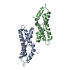
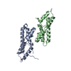
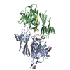
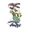
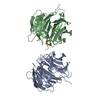
| Title | Crystal structure of red abalone egg VERL repeat 2 with linker in complex with sperm lysin at 1.8 A resolution | |||||||||||||||||||||
 Components Components |
| |||||||||||||||||||||
 Keywords Keywords | CELL ADHESION / FERTILIZATION / EGG-SPERM INTERACTION / GAMETE RECOGNITION / SPERM RECEPTOR / EGG COAT / VITELLINE ENVELOPE / ZP DOMAIN / ZP-N DOMAIN / SPERM ACROSOME / SPERM PENETRATION | |||||||||||||||||||||
| Function / homology |  Function and homology information Function and homology informationvitelline envelope / acrosomal lumen / sperm-egg recognition / single fertilization / plasma membrane Similarity search - Function | |||||||||||||||||||||
| Biological species |  Haliotis rufescens (red abalone) Haliotis rufescens (red abalone) | |||||||||||||||||||||
| Method |  X-RAY DIFFRACTION / SYNCHROTRON / MOLECULAR REPLACEMENT / Resolution: 1.8 Å X-RAY DIFFRACTION / SYNCHROTRON / MOLECULAR REPLACEMENT / Resolution: 1.8 Å | |||||||||||||||||||||
 Authors Authors | Nishimura, K. / Raj, I. / Sadat Al-Hosseini, H. / De Sanctis, D. / Jovine, L. | |||||||||||||||||||||
| Funding support |  Sweden, 6items Sweden, 6items
| |||||||||||||||||||||
 Citation Citation | Journal: Cell / Year: 2017 Title: Structural Basis of Egg Coat-Sperm Recognition at Fertilization. Authors: Raj, I. / Sadat Al Hosseini, H. / Dioguardi, E. / Nishimura, K. / Han, L. / Villa, A. / de Sanctis, D. / Jovine, L. #1: Journal: Mol. Biol. Evol. / Year: 2011 Title: The molecular basis of sex: linking yeast to human. Authors: Swanson, W.J. / Aagaard, J.E. / Vacquier, V.D. / Monne, M. / Sadat Al Hosseini, H. / Jovine, L. #2: Journal: Proc. Natl. Acad. Sci. U.S.A. / Year: 2006 Title: Rapidly evolving zona pellucida domain proteins are a major component of the vitelline envelope of abalone eggs. Authors: Aagaard, J.E. / Yi, X. / MacCoss, M.J. / Swanson, W.J. #3: Journal: Gene / Year: 2002 Title: Full-length sequence of VERL, the egg vitelline envelope receptor for abalone sperm lysin. Authors: Galindo, B.E. / Moy, G.W. / Swanson, W.J. / Vacquier, V.D. #4: Journal: Acta Crystallogr. D Biol. Crystallogr. / Year: 2000Title: 1.35 and 2.07 A resolution structures of the red abalone sperm lysin monomer and dimer reveal features involved in receptor binding. Authors: Kresge, N. / Vacquier, V.D. / Stout, C.D. #5: Journal: Proc. Natl. Acad. Sci. U.S.A. / Year: 1997 Title: The abalone egg vitelline envelope receptor for sperm lysin is a giant multivalent molecule. Authors: Swanson, W.J. / Vacquier, V.D. #6: Journal: J. Cell Biol. / Year: 1995Title: Crystal structure and subunit dynamics of the abalone sperm lysin dimer: egg envelopes dissociate dimers, the monomer is the active species. Authors: Shaw, A. / Fortes, P.A. / Stout, C.D. / Vacquier, V.D. #7: Journal: Science / Year: 1993Title: The crystal structure of lysin, a fertilization protein. Authors: Shaw, A. / McRee, D.E. / Vacquier, V.D. / Stout, C.D. | |||||||||||||||||||||
| History |
|
- Structure visualization
| Structure viewer | Molecule: MolmilJmol/JSmol |
|---|
- Downloads & links
-Download
| PDBx/mmCIF format | 5mr3.cif.gz | 597.8 KB | Display | PDBx/mmCIF format |
|---|---|---|---|---|
| PDB format | pdb5mr3.ent.gz | 502.2 KB | Display | PDB format |
| PDBx/mmJSON format | 5mr3.json.gz | Tree view | PDBx/mmJSON format | |
| Others |  Other downloads Other downloads |
-Validation report
| Arichive directory | https://data.pdbj.org/pub/pdb/validation_reports/mr/5mr3ftp://data.pdbj.org/pub/pdb/validation_reports/mr/5mr3 | HTTPS FTP |
|---|
-Related structure data
| Related structure data | 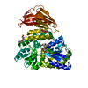 5ii4C 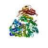 5ii5C  5ii6C 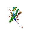 5ii7C 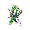 5ii8SC 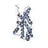 5ii9SC  5iiaC 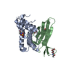 5iibC 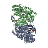 5iicC  5mr2C C: citing same article ( S: Starting model for refinement |
|---|---|
| Similar structure data |


-Links
 PDBj
PDBj- Assembly
| Deposited unit | 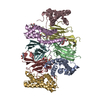
| ||||||||
|---|---|---|---|---|---|---|---|---|---|
| 1 | 
| ||||||||
| 2 | 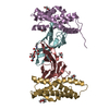
| ||||||||
| Unit cell |
|
-Components
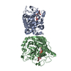
-Protein , 2 types, 8 molecules ACEGBDFH
| #1: Protein | Mass: 16295.218 Da / Num. of mol.: 4 Source method: isolated from a genetically manipulated source Source: (gene. exp.) Haliotis rufescens (red abalone) / Production host:  Homo sapiens (human) / References: UniProt: P04552 Homo sapiens (human) / References: UniProt: P04552#2: Protein | Mass: 15089.334 Da / Num. of mol.: 4 / Mutation: S293A, S296A, S297A Source method: isolated from a genetically manipulated source Source: (gene. exp.) Haliotis rufescens (red abalone) / Gene: VERL / Plasmid: pHLsec / Cell line (production host): HEK-293S / Production host: Homo sapiens (human) / References: UniProt: Q8WR62 |
|---|
-Sugars , 1 types, 4 molecules 
| #5: Sugar | ChemComp-NAG /  Type: D-saccharide, beta linking / Mass: 221.208 Da / Num. of mol.: 4 / Mutation: S293A, S296A, S297A Type: D-saccharide, beta linking / Mass: 221.208 Da / Num. of mol.: 4 / Mutation: S293A, S296A, S297ASource method: isolated from a genetically manipulated source Formula: C8H15NO6 / Source: (gene. exp.) Haliotis rufescens (red abalone) / Gene: VERL / Plasmid: pHLsec / Cell line (production host): HEK-293S / Production host: Homo sapiens (human) |
|---|
-Non-polymers , 4 types, 575 molecules 


| #3: Chemical | ChemComp-GOL /  Mass: 92.094 Da / Num. of mol.: 12 / Source method: obtained synthetically / Formula: C3H8O3 Mass: 92.094 Da / Num. of mol.: 12 / Source method: obtained synthetically / Formula: C3H8O3#4: Chemical |  Mass: 35.453 Da / Num. of mol.: 2 / Source method: obtained synthetically / Formula: Cl Mass: 35.453 Da / Num. of mol.: 2 / Source method: obtained synthetically / Formula: Cl#6: Chemical |  Mass: 150.173 Da / Num. of mol.: 2 / Source method: obtained synthetically / Formula: C6H14O4 Mass: 150.173 Da / Num. of mol.: 2 / Source method: obtained synthetically / Formula: C6H14O4#7: Water | ChemComp-HOH / | Mass: 18.015 Da / Num. of mol.: 559 / Source method: isolated from a natural source / Formula: H2O |
|---|
-Details
| Has protein modification | Y |
|---|
-Experimental details
-Experiment
| Experiment | Method: X-RAY DIFFRACTION / Number of used crystals: 1 |
|---|
- Sample preparation
| Crystal | Density Matthews: 2.62 Å3/Da / Density % sol: 53.09 % |
|---|---|
| Crystal grow | Temperature: 293 K / Method: vapor diffusion, sitting drop / pH: 8.5 Details: 5% (w/v) PEG 8000, 20% (v/v) PEG 300, 10% (v/v) Glycerol, 0.1 M Tris pH 8.5 |
-Data collection
| Diffraction | Mean temperature: 100 K |
|---|---|
| Diffraction source | Source: SYNCHROTRON / Site: ESRF  / Beamline: ID29 / Wavelength: 1.07227 Å / Beamline: ID29 / Wavelength: 1.07227 Å |
| Detector | Type: DECTRIS PILATUS 6M-F / Detector: PIXEL / Date: Sep 2, 2016 |
| Radiation | Protocol: SINGLE WAVELENGTH / Monochromatic (M) / Laue (L): M / Scattering type: x-ray |
| Radiation wavelength | Wavelength: 1.07227 Å / Relative weight: 1 |
| Reflection | Resolution: 1.8→45.322 Å / Num. obs: 109995 / % possible obs: 99.5 % / Redundancy: 3.4 % / CC1/2: 0.998 / Rmerge(I) obs: 0.05377 / Net I/σ(I): 11.53 |
| Reflection shell | Resolution: 1.8→1.864 Å / Redundancy: 3.4 % / Rmerge(I) obs: 1.494 / Mean I/σ(I) obs: 0.97 / CC1/2: 0.307 / % possible all: 99.6 |
- Processing
| Software |
| |||||||||||||||||||||||||||||||||||||||||||||||||||||||||||||||||||||||||||||||||||||||||||||||||||||||||||||||||||||||||||||||||||||||||||||||||||||||||||||||||||||||||||||||||||||||||||||||||||||||||||||||||||||||||||||||||
|---|---|---|---|---|---|---|---|---|---|---|---|---|---|---|---|---|---|---|---|---|---|---|---|---|---|---|---|---|---|---|---|---|---|---|---|---|---|---|---|---|---|---|---|---|---|---|---|---|---|---|---|---|---|---|---|---|---|---|---|---|---|---|---|---|---|---|---|---|---|---|---|---|---|---|---|---|---|---|---|---|---|---|---|---|---|---|---|---|---|---|---|---|---|---|---|---|---|---|---|---|---|---|---|---|---|---|---|---|---|---|---|---|---|---|---|---|---|---|---|---|---|---|---|---|---|---|---|---|---|---|---|---|---|---|---|---|---|---|---|---|---|---|---|---|---|---|---|---|---|---|---|---|---|---|---|---|---|---|---|---|---|---|---|---|---|---|---|---|---|---|---|---|---|---|---|---|---|---|---|---|---|---|---|---|---|---|---|---|---|---|---|---|---|---|---|---|---|---|---|---|---|---|---|---|---|---|---|---|---|---|---|---|---|---|---|---|---|---|---|---|---|---|---|---|---|---|
| Refinement | Method to determine structure: MOLECULAR REPLACEMENT Starting model: PDB ID 5II8, 5II9, D_1200000737 Resolution: 1.8→45.322 Å / SU ML: 0.31 / Cross valid method: FREE R-VALUE / σ(F): 1.33 / Phase error: 28.68
| |||||||||||||||||||||||||||||||||||||||||||||||||||||||||||||||||||||||||||||||||||||||||||||||||||||||||||||||||||||||||||||||||||||||||||||||||||||||||||||||||||||||||||||||||||||||||||||||||||||||||||||||||||||||||||||||||
| Solvent computation | Shrinkage radii: 0.9 Å / VDW probe radii: 1.11 Å | |||||||||||||||||||||||||||||||||||||||||||||||||||||||||||||||||||||||||||||||||||||||||||||||||||||||||||||||||||||||||||||||||||||||||||||||||||||||||||||||||||||||||||||||||||||||||||||||||||||||||||||||||||||||||||||||||
| Refinement step | Cycle: LAST / Resolution: 1.8→45.322 Å
| |||||||||||||||||||||||||||||||||||||||||||||||||||||||||||||||||||||||||||||||||||||||||||||||||||||||||||||||||||||||||||||||||||||||||||||||||||||||||||||||||||||||||||||||||||||||||||||||||||||||||||||||||||||||||||||||||
| Refine LS restraints |
| |||||||||||||||||||||||||||||||||||||||||||||||||||||||||||||||||||||||||||||||||||||||||||||||||||||||||||||||||||||||||||||||||||||||||||||||||||||||||||||||||||||||||||||||||||||||||||||||||||||||||||||||||||||||||||||||||
| LS refinement shell |
| |||||||||||||||||||||||||||||||||||||||||||||||||||||||||||||||||||||||||||||||||||||||||||||||||||||||||||||||||||||||||||||||||||||||||||||||||||||||||||||||||||||||||||||||||||||||||||||||||||||||||||||||||||||||||||||||||
| Refinement TLS params. | Method: refined / Refine-ID: X-RAY DIFFRACTION
| |||||||||||||||||||||||||||||||||||||||||||||||||||||||||||||||||||||||||||||||||||||||||||||||||||||||||||||||||||||||||||||||||||||||||||||||||||||||||||||||||||||||||||||||||||||||||||||||||||||||||||||||||||||||||||||||||
| Refinement TLS group |
|
