 Movie
Movie Controller

[English] 日本語
 Yorodumi
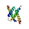
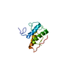
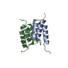
- PDB-5ii6: Crystal structure of the ZP-N1 domain of mouse sperm receptor ZP2... -
+ Open data

- Basic information
| Entry | Database: PDB / ID: 5ii6 | |||||||||||||||||||||
|---|---|---|---|---|---|---|---|---|---|---|---|---|---|---|---|---|---|---|---|---|---|---|
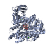
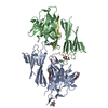
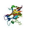
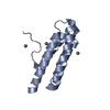
| Title | Crystal structure of the ZP-N1 domain of mouse sperm receptor ZP2 at 0.95 A resolution | |||||||||||||||||||||
 Components Components | Zona pellucida sperm-binding protein 2 | |||||||||||||||||||||
 Keywords Keywords | CELL ADHESION / FERTILIZATION / EGG-SPERM INTERACTION / GAMETE RECOGNITION / SPERM RECEPTOR / EGG COAT / ZONA PELLUCIDA / ZP DOMAIN / ZP-N DOMAIN | |||||||||||||||||||||
| Function / homology |  Function and homology information Function and homology informationInteraction With Cumulus Cells And The Zona Pellucida / structural constituent of egg coat / acrosin binding / egg coat / prevention of polyspermy / binding of sperm to zona pellucida / multivesicular body / extracellular matrix / endoplasmic reticulum / identical protein binding / plasma membrane Similarity search - Function | |||||||||||||||||||||
| Biological species |  | |||||||||||||||||||||
| Method |  X-RAY DIFFRACTION / SYNCHROTRON / SAD / Resolution: 0.95 Å X-RAY DIFFRACTION / SYNCHROTRON / SAD / Resolution: 0.95 Å | |||||||||||||||||||||
 Authors Authors | Dioguardi, E. / Han, L. / Nishimura, K. / De Sanctis, D. / Jovine, L. | |||||||||||||||||||||
| Funding support |  Sweden, 6items Sweden, 6items
| |||||||||||||||||||||
 Citation Citation | Journal: Cell / Year: 2017 Title: Structural Basis of Egg Coat-Sperm Recognition at Fertilization. Authors: Raj, I. / Sadat Al Hosseini, H. / Dioguardi, E. / Nishimura, K. / Han, L. / Villa, A. / de Sanctis, D. / Jovine, L. #1: Journal: J. Cell Biol. / Year: 2014 Title: A single domain of the ZP2 zona pellucida protein mediates gamete recognition in mice and humans. Authors: Avella, M.A. / Baibakov, B. / Dean, J. #2: Journal: J. Cell Biol. / Year: 2012 Title: Human sperm bind to the N-terminal domain of ZP2 in humanized zonae pellucidae in transgenic mice. Authors: Baibakov, B. / Boggs, N.A. / Yauger, B. / Baibakov, G. / Dean, J. #3: Journal: Science / Year: 2010 Title: Gamete recognition in mice depends on the cleavage status of an egg's zona pellucida protein. Authors: Gahlay, G. / Gauthier, L. / Baibakov, B. / Epifano, O. / Dean, J. #4: Journal: Nature / Year: 2008Title: Crystal structure of the ZP-N domain of ZP3 reveals the core fold of animal egg coats. Authors: Monne, M. / Han, L. / Schwend, T. / Burendahl, S. / Jovine, L. #5: Journal: J. Biol. Chem. / Year: 2003 Title: Structural characterization of native mouse zona pellucida proteins using mass spectrometry. Authors: Boja, E.S. / Hoodbhoy, T. / Fales, H.M. / Dean, J. #6: Journal: Mol. Cell. Biol. / Year: 1990 Title: Oocyte-specific expression of mouse Zp-2: developmental regulation of the zona pellucida genes. Authors: Liang, L.F. / Chamow, S.M. / Dean, J. #7: Journal: Cell / Year: 1982 Title: Biosynthesis of the major zona pellucida glycoprotein secreted by oocytes during mammalian oogenesis. Authors: Greve, J.M. / Salzmann, G.S. / Roller, R.J. / Wassarman, P.M. #8: Journal: Dev. Biol. / Year: 1981 Title: Mammalian sperm-egg interaction: fertilization of mouse eggs triggers modification of the major zona pellucida glycoprotein, ZP2. Authors: Bleil, J.D. / Beall, C.F. / Wassarman, P.M. #9: Journal: Proc. Natl. Acad. Sci. U.S.A. / Year: 1980 Title: Synthesis of zona pellucida proteins by denuded and follicle-enclosed mouse oocytes during culture in vitro. Authors: Bleil, J.D. / Wassarman, P.M. | |||||||||||||||||||||
| History |
|
- Structure visualization
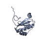
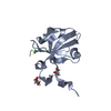
| Structure viewer | Molecule: MolmilJmol/JSmol |
|---|
- Downloads & links
-Download
| PDBx/mmCIF format | 5ii6.cif.gz | 85.9 KB | Display | PDBx/mmCIF format |
|---|---|---|---|---|
| PDB format | pdb5ii6.ent.gz | 65.5 KB | Display | PDB format |
| PDBx/mmJSON format | 5ii6.json.gz | Tree view | PDBx/mmJSON format | |
| Others |  Other downloads Other downloads |
-Validation report
| Arichive directory | https://data.pdbj.org/pub/pdb/validation_reports/ii/5ii6ftp://data.pdbj.org/pub/pdb/validation_reports/ii/5ii6 | HTTPS FTP |
|---|
-Related structure data
| Related structure data | 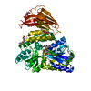 5ii4C 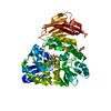 5ii5C 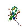 5ii7C 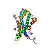 5ii8C 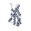 5ii9C  5iiaC  5iibC  5iicC  5mr2C  5mr3C C: citing same article ( |
|---|---|
| Similar structure data |


-Links
 PDBj
PDBj- Assembly
| Deposited unit | 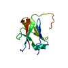
| ||||||||
|---|---|---|---|---|---|---|---|---|---|
| 1 |
| ||||||||
| Unit cell |
|
-Components
| #1: Protein | Mass: 12994.641 Da / Num. of mol.: 1 / Fragment: UNP residues 35-138 / Mutation: N83S Source method: isolated from a genetically manipulated source Source: (gene. exp.)  Homo sapiens (human) / References: UniProt: P20239 Homo sapiens (human) / References: UniProt: P20239 |
|---|---|
| #2: Water | ChemComp-HOH /  Mass: 18.015 Da / Num. of mol.: 142 / Source method: isolated from a natural source / Formula: H2O Mass: 18.015 Da / Num. of mol.: 142 / Source method: isolated from a natural source / Formula: H2O |
| Has protein modification | Y |
-Experimental details
-Experiment
| Experiment | Method: X-RAY DIFFRACTION / Number of used crystals: 1 |
|---|
- Sample preparation
| Crystal | Density Matthews: 2.15 Å3/Da / Density % sol: 42.8 % |
|---|---|
| Crystal grow | Temperature: 293 K / Method: vapor diffusion, hanging drop / pH: 6 / Details: 30% PEG 200, 5% PEG 3000, 0.1 M MES |
-Data collection
| Diffraction | Mean temperature: 100 K |
|---|---|
| Diffraction source | Source: SYNCHROTRON / Site: ESRF  / Beamline: ID29 / Wavelength: 0.8856 Å / Beamline: ID29 / Wavelength: 0.8856 Å |
| Detector | Type: DECTRIS PILATUS 6M-F / Detector: PIXEL / Date: Jul 25, 2012 |
| Radiation | Protocol: SINGLE WAVELENGTH / Monochromatic (M) / Laue (L): M / Scattering type: x-ray |
| Radiation wavelength | Wavelength: 0.8856 Å / Relative weight: 1 |
| Reflection | Resolution: 0.95→31.419 Å / Num. obs: 64506 / % possible obs: 99 % / Redundancy: 2 % / Biso Wilson estimate: 11.02 Å2 / CC1/2: 0.999 / Rmerge(I) obs: 0.02234 / Net I/σ(I): 13 |
| Reflection shell | Resolution: 0.95→0.984 Å / Redundancy: 1.9 % / Rmerge(I) obs: 0.2419 / Mean I/σ(I) obs: 2.66 / % possible all: 90 |
- Processing
| Software |
| ||||||||||||||||||||||||||||||||||||||||||||||||||||||||||||||||||||||||||||||||||||||||||||||||||||||||||||||||||||||||||||||||||||||||||||||||||||||||||||||||||||||||
|---|---|---|---|---|---|---|---|---|---|---|---|---|---|---|---|---|---|---|---|---|---|---|---|---|---|---|---|---|---|---|---|---|---|---|---|---|---|---|---|---|---|---|---|---|---|---|---|---|---|---|---|---|---|---|---|---|---|---|---|---|---|---|---|---|---|---|---|---|---|---|---|---|---|---|---|---|---|---|---|---|---|---|---|---|---|---|---|---|---|---|---|---|---|---|---|---|---|---|---|---|---|---|---|---|---|---|---|---|---|---|---|---|---|---|---|---|---|---|---|---|---|---|---|---|---|---|---|---|---|---|---|---|---|---|---|---|---|---|---|---|---|---|---|---|---|---|---|---|---|---|---|---|---|---|---|---|---|---|---|---|---|---|---|---|---|---|---|---|---|
| Refinement | Method to determine structure: SAD / Resolution: 0.95→31.419 Å / SU ML: 0.06 / Cross valid method: FREE R-VALUE / σ(F): 1.38 / Phase error: 12.12 / Stereochemistry target values: ML
| ||||||||||||||||||||||||||||||||||||||||||||||||||||||||||||||||||||||||||||||||||||||||||||||||||||||||||||||||||||||||||||||||||||||||||||||||||||||||||||||||||||||||
| Solvent computation | Shrinkage radii: 0.8 Å / VDW probe radii: 1.1 Å / Solvent model: FLAT BULK SOLVENT MODEL | ||||||||||||||||||||||||||||||||||||||||||||||||||||||||||||||||||||||||||||||||||||||||||||||||||||||||||||||||||||||||||||||||||||||||||||||||||||||||||||||||||||||||
| Refinement step | Cycle: LAST / Resolution: 0.95→31.419 Å
| ||||||||||||||||||||||||||||||||||||||||||||||||||||||||||||||||||||||||||||||||||||||||||||||||||||||||||||||||||||||||||||||||||||||||||||||||||||||||||||||||||||||||
| Refine LS restraints |
| ||||||||||||||||||||||||||||||||||||||||||||||||||||||||||||||||||||||||||||||||||||||||||||||||||||||||||||||||||||||||||||||||||||||||||||||||||||||||||||||||||||||||
| LS refinement shell |
|
