 Movie
Movie Controller

[English] 日本語
 Yorodumi
- PDB-2krd: Solution Structure of the Regulatory Domain of Human Cardiac Trop... -
+ Open data

- Basic information
| Entry | Database: PDB / ID: 2krd | ||||||
|---|---|---|---|---|---|---|---|
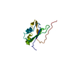
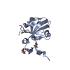
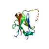
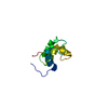
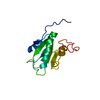
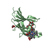
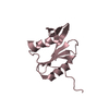
| Title | Solution Structure of the Regulatory Domain of Human Cardiac Troponin C in Complex with the Switch Region of cardiac Troponin I and W7 | ||||||
 Components Components |
| ||||||
 Keywords Keywords | STRUCTURAL PROTEIN / cardiac troponin C / regulatory domain / troponin I / switch region / W7 / Acetylation / Calcium / Cardiomyopathy / Disease mutation / Muscle protein / Polymorphism / Actin-binding / Phosphoprotein | ||||||
| Function / homology |  Function and homology information Function and homology informationregulation of systemic arterial blood pressure by ischemic conditions / troponin C binding / diaphragm contraction / regulation of muscle filament sliding speed / troponin T binding / cardiac Troponin complex / cardiac myofibril / negative regulation of ATP-dependent activity / troponin complex / regulation of muscle contraction ...regulation of systemic arterial blood pressure by ischemic conditions / troponin C binding / diaphragm contraction / regulation of muscle filament sliding speed / troponin T binding / cardiac Troponin complex / cardiac myofibril / negative regulation of ATP-dependent activity / troponin complex / regulation of muscle contraction / regulation of smooth muscle contraction / transition between fast and slow fiber / Striated Muscle Contraction / muscle filament sliding / regulation of cardiac muscle contraction by calcium ion signaling / response to metal ion / cardiac muscle cell contraction / ventricular cardiac muscle tissue morphogenesis / heart contraction / troponin I binding / vasculogenesis / calcium channel inhibitor activity / skeletal muscle contraction / Ion homeostasis / cardiac muscle contraction / sarcomere / intracellular calcium ion homeostasis / calcium-dependent protein binding / actin filament binding / heart development / actin binding / protein domain specific binding / calcium ion binding / protein kinase binding / protein homodimerization activity / cytosol Similarity search - Function | ||||||
| Biological species |  Homo sapiens (human) Homo sapiens (human) | ||||||
| Method | SOLUTION NMR / simulated annealing | ||||||
 Authors Authors | Oleszczuk, M. / Robertson, I.M. / Li, M.X. / Sykes, B.D. | ||||||
 Citation Citation | Journal: J.MOL.CELL.CARDIOL. / Year: 2010 Title: Solution structure of the regulatory domain of human cardiac troponin C in complex with the switch region of cardiac troponin I and W7: the basis of W7 as an inhibitor of cardiac muscle contraction. Authors: Oleszczuk, M. / Robertson, I.M. / Li, M.X. / Sykes, B.D. | ||||||
| History |
|
- Structure visualization
| Structure viewer | Molecule: MolmilJmol/JSmol |
|---|
- Downloads & links
-Download
| PDBx/mmCIF format | 2krd.cif.gz | 658.3 KB | Display | PDBx/mmCIF format |
|---|---|---|---|---|
| PDB format | pdb2krd.ent.gz | 551.6 KB | Display | PDB format |
| PDBx/mmJSON format | 2krd.json.gz | Tree view | PDBx/mmJSON format | |
| Others |  Other downloads Other downloads |
-Validation report
| Arichive directory | https://data.pdbj.org/pub/pdb/validation_reports/kr/2krdftp://data.pdbj.org/pub/pdb/validation_reports/kr/2krd | HTTPS FTP |
|---|
-Related structure data
| Similar structure data |
|---|


-Links
 PDBj
PDBj


- Assembly
| Deposited unit | 
| |||||||||
|---|---|---|---|---|---|---|---|---|---|---|
| 1 |
| |||||||||
| NMR ensembles |
|
-Components
| #1: Protein | Mass: 10070.304 Da / Num. of mol.: 1 Source method: isolated from a genetically manipulated source Source: (gene. exp.) Homo sapiens (human) / Gene: TNNC1, TNNC / Plasmid: pET3a / Production host:  |
|---|---|
| #2: Protein/peptide | Mass: 1806.183 Da / Num. of mol.: 1 / Source method: obtained synthetically / Source: (synth.) Homo sapiens (human) / References: UniProt: P19429 |
| #3: Chemical | ChemComp-CA /   Mass: 40.078 Da / Num. of mol.: 1 / Source method: obtained synthetically / Formula: Ca Mass: 40.078 Da / Num. of mol.: 1 / Source method: obtained synthetically / Formula: Ca |
| #4: Chemical | ChemComp-WW7 /   Mass: 340.868 Da / Num. of mol.: 1 / Source method: obtained synthetically / Formula: C16H21ClN2O2S Mass: 340.868 Da / Num. of mol.: 1 / Source method: obtained synthetically / Formula: C16H21ClN2O2S |
-Experimental details
-Experiment
| Experiment | Method: SOLUTION NMR | ||||||||||||||||||||||||||||||||||||||||||||||||||||||||||||
|---|---|---|---|---|---|---|---|---|---|---|---|---|---|---|---|---|---|---|---|---|---|---|---|---|---|---|---|---|---|---|---|---|---|---|---|---|---|---|---|---|---|---|---|---|---|---|---|---|---|---|---|---|---|---|---|---|---|---|---|---|---|
| NMR experiment |
|
 HSQC
HSQC- Sample preparation
| Details |
| ||||||||||||||||||||||||||||||||||||||||||||||||||||||||||||||||||||||||||||||||||||||||||||||||||||||
|---|---|---|---|---|---|---|---|---|---|---|---|---|---|---|---|---|---|---|---|---|---|---|---|---|---|---|---|---|---|---|---|---|---|---|---|---|---|---|---|---|---|---|---|---|---|---|---|---|---|---|---|---|---|---|---|---|---|---|---|---|---|---|---|---|---|---|---|---|---|---|---|---|---|---|---|---|---|---|---|---|---|---|---|---|---|---|---|---|---|---|---|---|---|---|---|---|---|---|---|---|---|---|---|
| Sample |
|