 Movie
Movie Controller

[English] 日本語
 Yorodumi
- PDB-5m2v: Structure of GluK1 ligand-binding domain (S1S2) in complex with (... -
+ Open data

- Basic information
| Entry | Database: PDB / ID: 5m2v | ||||||
|---|---|---|---|---|---|---|---|
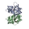
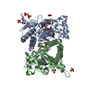
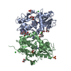
| Title | Structure of GluK1 ligand-binding domain (S1S2) in complex with (2S,4R)-4-(2-carboxyphenoxy)pyrrolidine-2-carboxylic acid at 3.18 A resolution | ||||||
 Components Components | Glutamate receptor ionotropic, kainate 1,Glutamate receptor ionotropic, kainate 1 | ||||||
 Keywords Keywords | MEMBRANE PROTEIN / Kainate receptor ligand-binding domain / GluK1-LBD / GluK1-S1S2 / antagonist | ||||||
| Function / homology |  Function and homology information Function and homology informationnegative regulation of synaptic transmission, GABAergic / gamma-aminobutyric acid secretion / L-glutamate transmembrane transporter activity / positive regulation of gamma-aminobutyric acid secretion / Activation of Na-permeable kainate receptors / kainate selective glutamate receptor complex / Activation of Ca-permeable Kainate Receptor / regulation of short-term neuronal synaptic plasticity / negative regulation of synaptic transmission, glutamatergic / glutamate binding ...negative regulation of synaptic transmission, GABAergic / gamma-aminobutyric acid secretion / L-glutamate transmembrane transporter activity / positive regulation of gamma-aminobutyric acid secretion / Activation of Na-permeable kainate receptors / kainate selective glutamate receptor complex / Activation of Ca-permeable Kainate Receptor / regulation of short-term neuronal synaptic plasticity / negative regulation of synaptic transmission, glutamatergic / glutamate binding / inhibitory postsynaptic potential / synaptic transmission, GABAergic / adult behavior / behavioral response to pain / modulation of excitatory postsynaptic potential / kainate selective glutamate receptor activity / extracellularly glutamate-gated ion channel activity / ionotropic glutamate receptor complex / membrane depolarization / glutamate-gated receptor activity / glutamate-gated calcium ion channel activity / ligand-gated monoatomic ion channel activity involved in regulation of presynaptic membrane potential / presynaptic modulation of chemical synaptic transmission / ionotropic glutamate receptor signaling pathway / SNARE binding / excitatory postsynaptic potential / regulation of membrane potential / positive regulation of synaptic transmission, GABAergic / transmitter-gated monoatomic ion channel activity involved in regulation of postsynaptic membrane potential / synaptic transmission, glutamatergic / establishment of localization in cell / postsynaptic density membrane / modulation of chemical synaptic transmission / regulation of synaptic plasticity / terminal bouton / nervous system development / presynaptic membrane / scaffold protein binding / chemical synaptic transmission / postsynaptic membrane / receptor complex / postsynaptic density / neuronal cell body / dendrite / synapse / glutamatergic synapse / identical protein binding / membrane / plasma membrane Similarity search - Function | ||||||
| Biological species |  | ||||||
| Method |  X-RAY DIFFRACTION / SYNCHROTRON / MOLECULAR REPLACEMENT / Resolution: 3.18 Å X-RAY DIFFRACTION / SYNCHROTRON / MOLECULAR REPLACEMENT / Resolution: 3.18 Å | ||||||
 Authors Authors | Frydenvang, K. / Kastrup, J.S. / Kristensen, C.M. | ||||||
 Citation Citation | Journal: J. Med. Chem. / Year: 2017 Title: Design and Synthesis of a Series of l-trans-4-Substituted Prolines as Selective Antagonists for the Ionotropic Glutamate Receptors Including Functional and X-ray Crystallographic Studies of ...Title: Design and Synthesis of a Series of l-trans-4-Substituted Prolines as Selective Antagonists for the Ionotropic Glutamate Receptors Including Functional and X-ray Crystallographic Studies of New Subtype Selective Kainic Acid Receptor Subtype 1 (GluK1) Antagonist (2S,4R)-4-(2-Carboxyphenoxy)pyrrolidine-2-carboxylic Acid. Authors: Krogsgaard-Larsen, N. / Delgar, C.G. / Koch, K. / Brown, P.M. / Moller, C. / Han, L. / Huynh, T.H. / Hansen, S.W. / Nielsen, B. / Bowie, D. / Pickering, D.S. / Kastrup, J.S. / Frydenvang, K. / Bunch, L. #1: Journal: FEBS Lett. / Year: 2005Title: Crystal structure of the kainate receptor GluR5 ligand-binding core in complex with (S)-glutamate. Authors: Naur, P. / Vestergaard, B. / Skov, L.K. / Egebjerg, J. / Gajhede, M. / Kastrup, J.S. | ||||||
| History |
|
- Structure visualization
| Structure viewer | Molecule: MolmilJmol/JSmol |
|---|
- Downloads & links
-Download
| PDBx/mmCIF format | 5m2v.cif.gz | 115.3 KB | Display | PDBx/mmCIF format |
|---|---|---|---|---|
| PDB format | pdb5m2v.ent.gz | 88.8 KB | Display | PDB format |
| PDBx/mmJSON format | 5m2v.json.gz | Tree view | PDBx/mmJSON format | |
| Others |  Other downloads Other downloads |
-Validation report
| Summary document | 5m2v_validation.pdf.gz | 985.3 KB | Display | wwPDB validaton report |
|---|---|---|---|---|
| Full document | 5m2v_full_validation.pdf.gz | 960.7 KB | Display | |
| Data in XML | 5m2v_validation.xml.gz | 19.5 KB | Display | |
| Data in CIF | 5m2v_validation.cif.gz | 25.4 KB | Display | |
| Arichive directory | https://data.pdbj.org/pub/pdb/validation_reports/m2/5m2vftp://data.pdbj.org/pub/pdb/validation_reports/m2/5m2v | HTTPS FTP |
-Related structure data
| Related structure data |  1vsoS S: Starting model for refinement |
|---|---|
| Similar structure data |


-Links
 PDBj
PDBj

- Assembly
| Deposited unit | 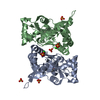
| ||||||||||||||||||||||||||||||||||||||||||||||||||||||||||||||||||||||||||||||||||||||
|---|---|---|---|---|---|---|---|---|---|---|---|---|---|---|---|---|---|---|---|---|---|---|---|---|---|---|---|---|---|---|---|---|---|---|---|---|---|---|---|---|---|---|---|---|---|---|---|---|---|---|---|---|---|---|---|---|---|---|---|---|---|---|---|---|---|---|---|---|---|---|---|---|---|---|---|---|---|---|---|---|---|---|---|---|---|---|---|
| 1 |
| ||||||||||||||||||||||||||||||||||||||||||||||||||||||||||||||||||||||||||||||||||||||
| Unit cell |
| ||||||||||||||||||||||||||||||||||||||||||||||||||||||||||||||||||||||||||||||||||||||
| Noncrystallographic symmetry (NCS) | NCS domain:
NCS domain segments: Ens-ID: 1 / Beg auth comp-ID: THR / Beg label comp-ID: THR
|