 Movie
Movie Controller

[English] 日本語
 Yorodumi
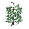
- PDB-1vso: Crystal Structure of the Ligand-Binding Core of iGluR5 in Complex... -
+ Open data

- Basic information
| Entry | Database: PDB / ID: 1vso | ||||||
|---|---|---|---|---|---|---|---|
| Title | Crystal Structure of the Ligand-Binding Core of iGluR5 in Complex With the Antagonist (S)-ATPO at 1.85 A resolution | ||||||
 Components Components | Glutamate receptor, ionotropic kainate 1 | ||||||
 Keywords Keywords | MEMBRANE PROTEIN / Antagonist complex | ||||||
| Function / homology |  Function and homology information Function and homology informationnegative regulation of synaptic transmission, GABAergic / L-glutamate transmembrane transporter activity / positive regulation of gamma-aminobutyric acid secretion / Activation of Na-permeable kainate receptors / Activation of Ca-permeable Kainate Receptor / kainate selective glutamate receptor complex / regulation of short-term neuronal synaptic plasticity / negative regulation of synaptic transmission, glutamatergic / glutamate binding / inhibitory postsynaptic potential ...negative regulation of synaptic transmission, GABAergic / L-glutamate transmembrane transporter activity / positive regulation of gamma-aminobutyric acid secretion / Activation of Na-permeable kainate receptors / Activation of Ca-permeable Kainate Receptor / kainate selective glutamate receptor complex / regulation of short-term neuronal synaptic plasticity / negative regulation of synaptic transmission, glutamatergic / glutamate binding / inhibitory postsynaptic potential / adult behavior / kainate selective glutamate receptor activity / behavioral response to pain / extracellularly glutamate-gated ion channel activity / modulation of excitatory postsynaptic potential / ionotropic glutamate receptor complex / membrane depolarization / glutamate-gated receptor activity / establishment of localization in cell / glutamate-gated calcium ion channel activity / ionotropic glutamate receptor signaling pathway / ligand-gated monoatomic ion channel activity involved in regulation of presynaptic membrane potential / presynaptic modulation of chemical synaptic transmission / positive regulation of synaptic transmission, GABAergic / SNARE binding / synaptic transmission, glutamatergic / transmitter-gated monoatomic ion channel activity involved in regulation of postsynaptic membrane potential / regulation of membrane potential / excitatory postsynaptic potential / regulation of synaptic plasticity / postsynaptic density membrane / modulation of chemical synaptic transmission / terminal bouton / nervous system development / presynaptic membrane / scaffold protein binding / chemical synaptic transmission / postsynaptic membrane / signaling receptor complex / postsynaptic density / neuronal cell body / synapse / dendrite / glutamatergic synapse / membrane / identical protein binding / plasma membrane Similarity search - Function | ||||||
| Biological species |  | ||||||
| Method |  X-RAY DIFFRACTION / SYNCHROTRON / MOLECULAR REPLACEMENT / Resolution: 1.85 Å X-RAY DIFFRACTION / SYNCHROTRON / MOLECULAR REPLACEMENT / Resolution: 1.85 Å | ||||||
 Authors Authors | Hald, H. / Naur, P. / Gajhede, M. / Kastrup, J.S. | ||||||
 Citation Citation | Journal: J.Biol.Chem. / Year: 2007 Title: Partial agonism and antagonism of the ionotropic glutamate receptor iGLuR5: structures of the ligand-binding core in complex with domoic acid and 2-amino-3-[5-tert-butyl-3-(phosphonomethoxy)-4- ...Title: Partial agonism and antagonism of the ionotropic glutamate receptor iGLuR5: structures of the ligand-binding core in complex with domoic acid and 2-amino-3-[5-tert-butyl-3-(phosphonomethoxy)-4-isoxazolyl]propionic acid. Authors: Hald, H. / Naur, P. / Pickering, D.S. / Sprogoe, D. / Madsen, U. / Timmermann, D.B. / Ahring, P.K. / Liljefors, T. / Schousboe, A. / Egebjerg, J. / Gajhede, M. / Kastrup, J.S. #1: Journal: FEBS Lett. / Year: 2005Title: Crystal structure of the kainate receptor GluR5 ligand-binding core in complex with (S)-glutamate. Authors: Naur, P. / Vestergaard, B. / Skov, L.K. / Egebjerg, J. / Gajhede, M. / Kastrup, J.S. #2: Journal: J.Neurosci. / Year: 2006Title: Crystal structures of the kainate receptor GluR5 ligand binding core dimer with novel GluR5-selective antagonists. Authors: Mayer, M.L. / Ghosal, A. / Dolman, N.P. / Jane, D.E. #3: Journal: J.Med.Chem. / Year: 2003Title: Competitive antagonism of AMPA receptors by ligands of different classes: crystal structure of ATPO bound to the GluR2 ligand-binding core, in comparison with DNQX. Authors: Hogner, A. / Greenwood, J.R. / Liljefors, T. / Lunn, M.L. / Egebjerg, J. / Larsen, I.K. / Gouaux, E. / Kastrup, J.S. | ||||||
| History |
| ||||||
| Remark 999 | sequence There is a Ala -> Gly sequence conflict at residue 477 in the UniProt database. | ||||||
| Remark 300 | BIOMOLECULE: 1 THIS ENTRY CONTAINS THE CRYSTALLOGRAPHIC ASYMMETRIC UNIT WHICH CONSISTS OF 1 CHAIN(S) ...BIOMOLECULE: 1 THIS ENTRY CONTAINS THE CRYSTALLOGRAPHIC ASYMMETRIC UNIT WHICH CONSISTS OF 1 CHAIN(S). Authors state the functional receptor is a tetramer built of dimers-of-dimers. However, in the crystal only the dimer is present. |
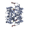
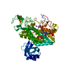
- Structure visualization
| Structure viewer | Molecule: MolmilJmol/JSmol |
|---|
- Downloads & links
-Download
| PDBx/mmCIF format | 1vso.cif.gz | 69.6 KB | Display | PDBx/mmCIF format |
|---|---|---|---|---|
| PDB format | pdb1vso.ent.gz | 49.5 KB | Display | PDB format |
| PDBx/mmJSON format | 1vso.json.gz | Tree view | PDBx/mmJSON format | |
| Others |  Other downloads Other downloads |
-Validation report
| Arichive directory | https://data.pdbj.org/pub/pdb/validation_reports/vs/1vsoftp://data.pdbj.org/pub/pdb/validation_reports/vs/1vso | HTTPS FTP |
|---|
-Related structure data
| Related structure data |  2pbwC  1n0tS S: Starting model for refinement C: citing same article ( |
|---|---|
| Similar structure data |


-Links
 PDBj
PDBj

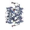
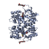
- Assembly
| Deposited unit | 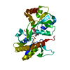
| |||||||||
|---|---|---|---|---|---|---|---|---|---|---|
| 1 | 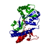
| |||||||||
| Unit cell |
| |||||||||
| Components on special symmetry positions |
|
-Components
| #1: Protein | Mass: 29108.453 Da / Num. of mol.: 1 Source method: isolated from a genetically manipulated source Source: (gene. exp.)  |
|---|---|
| #2: Chemical | ChemComp-AT1 / (  Mass: 322.252 Da / Num. of mol.: 1 / Source method: obtained synthetically / Formula: C11H19N2O7P Mass: 322.252 Da / Num. of mol.: 1 / Source method: obtained synthetically / Formula: C11H19N2O7P |
| #3: Chemical | ChemComp-GOL /   Mass: 92.094 Da / Num. of mol.: 1 / Source method: obtained synthetically / Formula: C3H8O3 Mass: 92.094 Da / Num. of mol.: 1 / Source method: obtained synthetically / Formula: C3H8O3 |
| #4: Water | ChemComp-HOH /  Mass: 18.015 Da / Num. of mol.: 204 / Source method: isolated from a natural source / Formula: H2O Mass: 18.015 Da / Num. of mol.: 204 / Source method: isolated from a natural source / Formula: H2O |
-Experimental details
-Experiment
| Experiment | Method: X-RAY DIFFRACTION / Number of used crystals: 1 |
|---|
- Sample preparation
| Crystal | Density Matthews: 3 Å3/Da / Density % sol: 59.05 % |
|---|---|
| Crystal grow | Temperature: 279 K / pH: 6.5 Details: 20% PEG 4000, 0.3 M lithium sulfate, 0.1 M cacodylate, VAPOR DIFFUSION, HANGING DROP, temperature 279K, pH 6.50 |
-Data collection
| Diffraction | Mean temperature: 100 K |
|---|---|
| Diffraction source | Source: SYNCHROTRON / Site: EMBL/DESY, HAMBURG  / Beamline: X11 / Wavelength: 0.812 / Beamline: X11 / Wavelength: 0.812 |
| Detector | Type: MAR CCD 165 mm / Detector: CCD / Date: Jun 13, 2005 |
| Radiation | Protocol: SINGLE WAVELENGTH / Monochromatic (M) / Laue (L): M / Scattering type: x-ray |
| Radiation wavelength | Wavelength: 0.812 Å / Relative weight: 1 |
| Reflection | Resolution: 1.85→25 Å / Num. obs: 29461 / % possible obs: 99.7 % / Observed criterion σ(I): -3 / Redundancy: 4.8 % / Biso Wilson estimate: 21.9 Å2 / Rsym value: 0.059 / Net I/σ(I): 23.1 |
| Reflection shell | Resolution: 1.85→1.92 Å / Mean I/σ(I) obs: 3.8 / Rsym value: 0.357 / % possible all: 99.9 |
-Phasing
| Phasing MR |
|
|---|
- Processing
| Software |
| ||||||||||||||||||||||||||||||||||||||||||||||||||||||||||||||||||||||||||||||||
|---|---|---|---|---|---|---|---|---|---|---|---|---|---|---|---|---|---|---|---|---|---|---|---|---|---|---|---|---|---|---|---|---|---|---|---|---|---|---|---|---|---|---|---|---|---|---|---|---|---|---|---|---|---|---|---|---|---|---|---|---|---|---|---|---|---|---|---|---|---|---|---|---|---|---|---|---|---|---|---|---|---|
| Refinement | Method to determine structure: MOLECULAR REPLACEMENT Starting model: PDB ENTRY 1N0T, CHAIN A Resolution: 1.85→24.66 Å / Rfactor Rfree error: 0.006 / Data cutoff high absF: 229797.41 / Data cutoff low absF: 0 / Isotropic thermal model: RESTRAINED / Cross valid method: THROUGHOUT / σ(F): 0 / Stereochemistry target values: ENGH & HUBER Details: THE FULLY REFINED STRUCTURE COMPRISES THR433-GLN492, TRP498-LYS544, THE GLY-THR LINKER, PRO667-SER711 AND SER715-GLY803
| ||||||||||||||||||||||||||||||||||||||||||||||||||||||||||||||||||||||||||||||||
| Solvent computation | Solvent model: FLAT MODEL / Bsol: 48.9 Å2 / ksol: 0.41 e/Å3 | ||||||||||||||||||||||||||||||||||||||||||||||||||||||||||||||||||||||||||||||||
| Displacement parameters | Biso mean: 24.7 Å2
| ||||||||||||||||||||||||||||||||||||||||||||||||||||||||||||||||||||||||||||||||
| Refine analyze |
| ||||||||||||||||||||||||||||||||||||||||||||||||||||||||||||||||||||||||||||||||
| Refinement step | Cycle: LAST / Resolution: 1.85→24.66 Å
| ||||||||||||||||||||||||||||||||||||||||||||||||||||||||||||||||||||||||||||||||
| Refine LS restraints |
| ||||||||||||||||||||||||||||||||||||||||||||||||||||||||||||||||||||||||||||||||
| LS refinement shell | Resolution: 1.85→1.93 Å / Rfactor Rfree error: 0.022 / Total num. of bins used: 8
| ||||||||||||||||||||||||||||||||||||||||||||||||||||||||||||||||||||||||||||||||
| Xplor file |
|
