[English] 日本語
Yorodumi
- PDB-3ng0: Crystal Structure of Glutamine Synthetase from Synechocystis sp. ... -

+
Open data


ID or keywords:

Loading...

-
Basic information

Entry
Database: PDB / ID: 3ng0
TitleCrystal Structure of Glutamine Synthetase from Synechocystis sp. PCC 6803
ComponentsGlutamine synthetase
KeywordsLIGASE / Glutamine synthetase type I / GSI / Nitrogen Metabolism / Synechocystis
Function / homology
Function and homology information


nitrogen utilization / glutamine synthetase / glutamine biosynthetic process / glutamine synthetase activity / ATP binding / membrane / metal ion binding / cytoplasm
Similarity search - Function
Glutamine synthetase, N-terminal domain / Glutamine synthetase type I / Glutamine synthetase/guanido kinase, catalytic domain / Glutamine synthetase, N-terminal conserved site / Glutamine synthetase signature 1. / Glutamine synthetase, beta-Grasp domain / Creatine Kinase; Chain A, domain 2 / Glutamine synthetase, glycine-rich site / Glutamine synthetase putative ATP-binding region signature. / Glutamine synthetase, N-terminal domain ...Glutamine synthetase, N-terminal domain / Glutamine synthetase type I / Glutamine synthetase/guanido kinase, catalytic domain / Glutamine synthetase, N-terminal conserved site / Glutamine synthetase signature 1. / Glutamine synthetase, beta-Grasp domain / Creatine Kinase; Chain A, domain 2 / Glutamine synthetase, glycine-rich site / Glutamine synthetase putative ATP-binding region signature. / Glutamine synthetase, N-terminal domain / Glutamine synthetase, N-terminal domain superfamily / Glutamine synthetase, catalytic domain / Glutamine synthetase, catalytic domain / Glutamine synthetase, catalytic domain / Glutamine synthetase/guanido kinase, catalytic domain / Ubiquitin-like (UB roll) / Roll / 2-Layer Sandwich / Alpha Beta
Similarity search - Domain/homology
PHOSPHOAMINOPHOSPHONIC ACID-ADENYLATE ESTER / : / Glutamine synthetase
Similarity search - Component
Biological speciesSynechocystis sp. (bacteria)
MethodX-RAY DIFFRACTION / SYNCHROTRON / MOLECULAR REPLACEMENT / molecular replacement / Resolution: 2.8 Å
AuthorsSaelices, L. / Cascio, D. / Florencio, F.J. / Muro-Pastor, M.I.
CitationJournal: To be Published
Title: Crystal Structure of Glutamine Synthetase from Synechocystis sp. PCC 6803
Authors: Saelices, L. / Cascio, D. / Florencio, F.J. / Muro-Pastor, M.I.
History
DepositionJun 10, 2010Deposition site: RCSB / Processing site: RCSB
Revision 1.0Jun 15, 2011Provider: repository / Type: Initial release
Revision 1.1Jul 13, 2011Group: Version format compliance
Revision 1.2Feb 21, 2024Group: Data collection / Database references / Derived calculations
Category: chem_comp_atom / chem_comp_bond ...chem_comp_atom / chem_comp_bond / database_2 / pdbx_struct_conn_angle / struct_conn / struct_ref_seq_dif / struct_site
Item: _database_2.pdbx_DOI / _database_2.pdbx_database_accession ..._database_2.pdbx_DOI / _database_2.pdbx_database_accession / _pdbx_struct_conn_angle.ptnr1_auth_comp_id / _pdbx_struct_conn_angle.ptnr1_auth_seq_id / _pdbx_struct_conn_angle.ptnr1_label_asym_id / _pdbx_struct_conn_angle.ptnr1_label_atom_id / _pdbx_struct_conn_angle.ptnr1_label_comp_id / _pdbx_struct_conn_angle.ptnr1_label_seq_id / _pdbx_struct_conn_angle.ptnr2_auth_seq_id / _pdbx_struct_conn_angle.ptnr2_label_asym_id / _pdbx_struct_conn_angle.ptnr3_auth_comp_id / _pdbx_struct_conn_angle.ptnr3_auth_seq_id / _pdbx_struct_conn_angle.ptnr3_label_asym_id / _pdbx_struct_conn_angle.ptnr3_label_atom_id / _pdbx_struct_conn_angle.ptnr3_label_comp_id / _pdbx_struct_conn_angle.ptnr3_label_seq_id / _pdbx_struct_conn_angle.value / _struct_conn.pdbx_dist_value / _struct_conn.ptnr1_auth_comp_id / _struct_conn.ptnr1_auth_seq_id / _struct_conn.ptnr1_label_asym_id / _struct_conn.ptnr1_label_atom_id / _struct_conn.ptnr1_label_comp_id / _struct_conn.ptnr1_label_seq_id / _struct_conn.ptnr2_auth_seq_id / _struct_conn.ptnr2_label_asym_id / _struct_ref_seq_dif.details / _struct_site.pdbx_auth_asym_id / _struct_site.pdbx_auth_comp_id / _struct_site.pdbx_auth_seq_id

-
Structure visualization

Structure viewerMolecule:
MolmilJmol/JSmol

Downloads & links

-
Assembly

Deposited unit
A: Glutamine synthetase
hetero molecules


Theoretical massNumber of molelcules
Total (without water)53,7255
Polymers53,0541
Non-polymers6714
Water43224
1
A: Glutamine synthetase
hetero molecules
x 12


Theoretical massNumber of molelcules
Total (without water)644,69560
Polymers636,64312
Non-polymers8,05248
Water21612
TypeNameSymmetry operationNumber
identity operation1_555x,y,z1
crystal symmetry operation2_555-y,x-y,z1
crystal symmetry operation3_555-x+y,-x,z1
crystal symmetry operation4_555-x,-y,z1
crystal symmetry operation5_555y,-x+y,z1
crystal symmetry operation6_555x-y,x,z1
crystal symmetry operation7_556y,x,-z+11
crystal symmetry operation8_556x-y,-y,-z+11
crystal symmetry operation9_556-x,-x+y,-z+11
crystal symmetry operation10_556-y,-x,-z+11
crystal symmetry operation11_556-x+y,y,-z+11
crystal symmetry operation12_556x,x-y,-z+11
Buried area99130 Å2
ΔGint-761 kcal/mol
Surface area173520 Å2
MethodPISA
Unit cell
Length a, b, c (Å)132.016, 132.016, 202.130
Angle α, β, γ (deg.)90.00, 90.00, 120.00
Int Tables number177
Space group name H-MP622

-
Components

#1: Protein Glutamine synthetase / / Glutamate--ammonia ligase


Mass: 53053.551 Da / Num. of mol.: 1
Source method: isolated from a genetically manipulated source
Source: (gene. exp.) Synechocystis sp. (bacteria) / Strain: PCC 6803 / Gene: glnA, slr1756 / Plasmid: pBS-SK+ / Production host: Escherichia coli (E. coli) / Strain (production host): DH5-ALPHA / References: UniProt: P77961, glutamine synthetase
#2: Chemical ChemComp-ANP / PHOSPHOAMINOPHOSPHONIC ACID-ADENYLATE ESTER


Mass: 506.196 Da / Num. of mol.: 1 / Source method: obtained synthetically / Formula: C10H17N6O12P3 / Comment: AMP-PNP, energy-carrying molecule analogue*YM
#3: Chemical ChemComp-MN / MANGANESE (II) ION


Mass: 54.938 Da / Num. of mol.: 3 / Source method: obtained synthetically / Formula: Mn
#4: Water ChemComp-HOH / water / Water


Mass: 18.015 Da / Num. of mol.: 24 / Source method: isolated from a natural source / Formula: H2O
Compound details
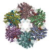
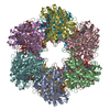
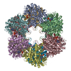
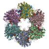
THIS CRYSTAL CONTAINS 2 MOLECULES PER ASYMMETRIC UNIT. P622 CRYSTAL SYMMETRY GENERATES TWO ...THIS CRYSTAL CONTAINS 2 MOLECULES PER ASYMMETRIC UNIT. P622 CRYSTAL SYMMETRY GENERATES TWO DODECAMERS (BIOLOGICAL UNITS) IN THE UNIT CELL, ONE DODECAMER FROM EACH MONOMER. UNIT CELL TRANSLATIONS ALONG A AND B GENERATE LAYERS IN THE CRYSTAL COMPOSED OF DODECAMER 1 WHICH ARE INTERLEAVED BY SIMILAR LAYERS COMPOSED OF DODECAMER 2. TRANSLOCATION LATTICE DEFECTS (TLD) COMPLICATE THE INTERPRETATION OF THE ELECTRON DENSITY FOR DODECAMER 2. THIS DODECAMER IS DISTRIBUTED RANDOMLY OVER 6 DIFFERENT POSITIONS (DIFFERING IN X,Y COORDINATES) WITHIN THE LAYER. TO FACILITATE INTERPRETATION OF THE ELECTRON DENSITY MAP, THE CONTRIBUTION FROM DODECAMER 2 HAS BEEN REMOVED FROM THE STRUCTURE FACTORS BY APPLYING A LATTICE TRANSLOCATION CORRECTION. ACCORDINGLY, THE COORDINATES FOR THE SECOND MOLECULE ARE NOT INCLUDED IN THIS PDB ENTRY.

-
Experimental details

-
Experiment

ExperimentMethod: X-RAY DIFFRACTION / Number of used crystals: 1

-
Sample preparation

CrystalDensity Matthews: 2.49 Å3/Da / Density % sol: 50.55 %
Crystal growTemperature: 298 K / Method: vapor diffusion, hanging drop / pH: 4
Details: 0.1M sodium acetate,0.1M calcium acetate, 10% PEG 4000, pH 4.0, vapor diffusion, hanging drop, temperature 298K

-
Data collection

DiffractionMean temperature: 100 K
Diffraction sourceSource: SYNCHROTRON / Site: APS / Beamline: 24-ID-C / Wavelength: 0.9791 Å
DetectorType: ADSC QUANTUM 315 / Detector: CCD / Date: Nov 12, 2009
RadiationMonochromator: Si (111) / Protocol: SINGLE WAVELENGTH / Monochromatic (M) / Laue (L): M / Scattering type: x-ray
Radiation wavelengthWavelength: 0.9791 Å / Relative weight: 1
ReflectionResolution: 2.8→100 Å / Num. obs: 25802 / % possible obs: 98 % / Redundancy: 6.7 % / Biso Wilson estimate: 54.56 Å2 / Rmerge(I) obs: 0.213 / Net I/σ(I): 4.1
Reflection shell
Resolution (Å)Redundancy (%)Rmerge(I) obsDiffraction-ID% possible all
2.8-2.96.80.424199.2
2.9-3.026.80.37199.5
3.02-3.156.80.302199.3
3.15-3.326.80.274199.1
3.32-3.536.90.233198.7
3.53-3.86.70.21198.7
3.8-4.186.80.196198.4
4.18-4.796.50.178197.5
4.79-6.036.70.176197.2
6.03-1006.30.157193.4

-
Phasing

PhasingMethod: molecular replacement
Phasing MRRfactor: 54.72 / Model details: Phaser MODE: MR_AUTO
Highest resolutionLowest resolution
Rotation2.8 Å99.51 Å
Translation2.8 Å99.51 Å

-
Processing

Software
NameVersionClassificationNB
SCALEPACKdata scaling
PHASER2.1.4phasing
BUSTER-TNTrefinement
PDB_EXTRACT3.1data extraction
BUSTER2.8.0refinement
RefinementMethod to determine structure: MOLECULAR REPLACEMENT / Resolution: 2.8→32.92 Å / Cor.coef. Fo:Fc: 0.8182 / Cor.coef. Fo:Fc free: 0.7756 / Occupancy max: 1 / Occupancy min: 0 / Cross valid method: THROUGHOUT / σ(F): 0
RfactorNum. reflection% reflectionSelection details
Rfree0.3129 1298 5.04 %RANDOM
Rwork0.2853 ---
obs0.2867 25779 --
Displacement parametersBiso mean: 28.98 Å2
Baniso -1Baniso -2Baniso -3
1-6.6465 Å20 Å20 Å2
2--6.6465 Å20 Å2
3----13.293 Å2
Refine analyzeLuzzati coordinate error obs: 0.546 Å
Refinement stepCycle: LAST / Resolution: 2.8→32.92 Å
ProteinNucleic acidLigandSolventTotal
Num. atoms3674 0 34 24 3732
Refine LS restraints
Refine-IDTypeDev idealNumberRestraint functionWeight
X-RAY DIFFRACTIONt_bond_d0.0093811HARMONIC2
X-RAY DIFFRACTIONt_angle_deg1.165180HARMONIC2
X-RAY DIFFRACTIONt_dihedral_angle_d1299SINUSOIDAL2
X-RAY DIFFRACTIONt_incorr_chiral_ct
X-RAY DIFFRACTIONt_pseud_angle
X-RAY DIFFRACTIONt_trig_c_planes97HARMONIC2
X-RAY DIFFRACTIONt_gen_planes546HARMONIC5
X-RAY DIFFRACTIONt_it3778HARMONIC20
X-RAY DIFFRACTIONt_nbd1SEMIHARMONIC5
X-RAY DIFFRACTIONt_omega_torsion3.48
X-RAY DIFFRACTIONt_other_torsion21.53
X-RAY DIFFRACTIONt_improper_torsion
X-RAY DIFFRACTIONt_chiral_improper_torsion4855
X-RAY DIFFRACTIONt_sum_occupancies
X-RAY DIFFRACTIONt_utility_distance
X-RAY DIFFRACTIONt_utility_angle
X-RAY DIFFRACTIONt_utility_torsion
X-RAY DIFFRACTIONt_ideal_dist_contact44734
LS refinement shellResolution: 2.8→2.91 Å / Total num. of bins used: 13
RfactorNum. reflection% reflection
Rfree0.3443 124 4.4 %
Rwork0.3031 2693 -
all0.3048 2817 -

+
About Yorodumi

-
News

-
Feb 9, 2022. New format data for meta-information of EMDB entries

New format data for meta-information of EMDB entries

  • Version 3 of the EMDB header file is now the official format.
  • The previous official version 1.9 will be removed from the archive.

Related info.:EMDB header

External links:wwPDB to switch to version 3 of the EMDB data model

-
Aug 12, 2020. Covid-19 info

Covid-19 info

URL: https://pdbj.org/emnavi/covid19.php

New page: Covid-19 featured information page in EM Navigator.

Related info.:Covid-19 info / Mar 5, 2020. Novel coronavirus structure data

+
Mar 5, 2020. Novel coronavirus structure data

Novel coronavirus structure data

Related info.:Yorodumi Speices / Aug 12, 2020. Covid-19 info

External links:COVID-19 featured content - PDBj / Molecule of the Month (242):Coronavirus Proteases

+
Jan 31, 2019. EMDB accession codes are about to change! (news from PDBe EMDB page)

EMDB accession codes are about to change! (news from PDBe EMDB page)

  • The allocation of 4 digits for EMDB accession codes will soon come to an end. Whilst these codes will remain in use, new EMDB accession codes will include an additional digit and will expand incrementally as the available range of codes is exhausted. The current 4-digit format prefixed with “EMD-” (i.e. EMD-XXXX) will advance to a 5-digit format (i.e. EMD-XXXXX), and so on. It is currently estimated that the 4-digit codes will be depleted around Spring 2019, at which point the 5-digit format will come into force.
  • The EM Navigator/Yorodumi systems omit the EMD- prefix.

Related info.:Q: What is EMD? / ID/Accession-code notation in Yorodumi/EM Navigator

External links:EMDB Accession Codes are Changing Soon! / Contact to PDBj

+
Jul 12, 2017. Major update of PDB

Major update of PDB

  • wwPDB released updated PDB data conforming to the new PDBx/mmCIF dictionary.
  • This is a major update changing the version number from 4 to 5, and with Remediation, in which all the entries are updated.
  • In this update, many items about electron microscopy experimental information are reorganized (e.g. em_software).
  • Now, EM Navigator and Yorodumi are based on the updated data.

External links:wwPDB Remediation / Enriched Model Files Conforming to OneDep Data Standards Now Available in the PDB FTP Archive

-
Yorodumi

Thousand views of thousand structures

  • Yorodumi is a browser for structure data from EMDB, PDB, SASBDB, etc.
  • This page is also the successor to EM Navigator detail page, and also detail information page/front-end page for Omokage search.
  • The word "yorodu" (or yorozu) is an old Japanese word meaning "ten thousand". "mi" (miru) is to see.

Related info.:EMDB / PDB / SASBDB / Comparison of 3 databanks / Yorodumi Search / Aug 31, 2016. New EM Navigator & Yorodumi / Yorodumi Papers / Jmol/JSmol / Function and homology information / Changes in new EM Navigator and Yorodumi

Read more