 Movie
Movie Controller

[English] 日本語
 Yorodumi
- PDB-2wgs: Crystal structure of Mycobacterium Tuberculosis Glutamine Synthet... -
+ Open data

- Basic information
| Entry | Database: PDB / ID: 2wgs | ||||||
|---|---|---|---|---|---|---|---|
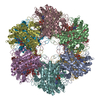
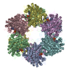
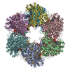
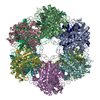
| Title | Crystal structure of Mycobacterium Tuberculosis Glutamine Synthetase in complex with a purine analogue inhibitor. | ||||||
 Components Components | GLUTAMINE SYNTHETASE 1 | ||||||
 Keywords Keywords | LIGASE / RELAXED STATE / PURINE ANALOGUE / NUCLEOTIDE-BINDING / GLNA1 / MT2278 / RV2220 / CYTOPLASM / SYNTHETASE | ||||||
| Function / homology |  Function and homology information Function and homology informationnitrogen utilization / positive regulation of plasminogen activation / cell wall / glutamine synthetase / : / glutamine synthetase activity / zymogen binding / cobalt ion binding / fibronectin binding / peptidoglycan-based cell wall ...nitrogen utilization / positive regulation of plasminogen activation / cell wall / glutamine synthetase / : / glutamine synthetase activity / zymogen binding / cobalt ion binding / fibronectin binding / peptidoglycan-based cell wall / ADP binding / protein homooligomerization / GDP binding / manganese ion binding / GTP binding / magnesium ion binding / extracellular region / ATP binding / membrane / metal ion binding / plasma membrane / cytoplasm / cytosol Similarity search - Function | ||||||
| Biological species |   MYCOBACTERIUM TUBERCULOSIS (bacteria) MYCOBACTERIUM TUBERCULOSIS (bacteria) | ||||||
| Method |  X-RAY DIFFRACTION / SYNCHROTRON / MOLECULAR REPLACEMENT / Resolution: 2.55 Å X-RAY DIFFRACTION / SYNCHROTRON / MOLECULAR REPLACEMENT / Resolution: 2.55 Å | ||||||
 Authors Authors | Nilsson, M.T. / Krajewski, W.W. / Jones, T.A. / Mowbray, S.L. | ||||||
 Citation Citation | Journal: J.Mol.Biol. / Year: 2009 Title: Structural Basis for the Inhibition of Mycobacterium Tuberculosis Glutamine Synthetase by Novel ATP-Competitive Inhibitors. Authors: Nilsson, M.T. / Krajewski, W.W. / Yellagunda, S. / Prabhumurthy, S. / Chamarahally, G.N. / Siddamadappa, C. / Srinivasa, B.R. / Yahiaoui, S. / Larhed, M. / Karlen, A. / Jones, T.A. / Mowbray, S.L. #1: Journal: Proc.Natl.Acad.Sci.USA / Year: 2005Title: Structure of Mycobacterium Tuberculosis Glutamine Synthetase in Complex with a Transition-State Mimic Provides Functional Insights. Authors: Krajewski, W.W. / Jones, T.A. / Mowbray, S.L. | ||||||
| History |
| ||||||
| Remark 650 | HELIX DETERMINATION METHOD: AUTHOR PROVIDED. | ||||||
| Remark 700 | SHEET DETERMINATION METHOD: AUTHOR PROVIDED. |
- Structure visualization
| Structure viewer | Molecule: MolmilJmol/JSmol |
|---|
- Downloads & links
-Download
| PDBx/mmCIF format | 2wgs.cif.gz | 1.1 MB | Display | PDBx/mmCIF format |
|---|---|---|---|---|
| PDB format | pdb2wgs.ent.gz | 904.1 KB | Display | PDB format |
| PDBx/mmJSON format | 2wgs.json.gz | Tree view | PDBx/mmJSON format | |
| Others |  Other downloads Other downloads |
-Validation report
| Arichive directory | https://data.pdbj.org/pub/pdb/validation_reports/wg/2wgsftp://data.pdbj.org/pub/pdb/validation_reports/wg/2wgs | HTTPS FTP |
|---|
-Related structure data
| Related structure data | 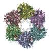 2whiC 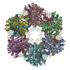 1htoS S: Starting model for refinement C: citing same article ( |
|---|---|
| Similar structure data |


-Links
 PDBj
PDBj
- Assembly
| Deposited unit | 
| |||||||||||||||||||||||||||||||||||||||||||||||||||||||||||||||||||||||||||||||||||||||||||||||||||||||||||||||||||||
|---|---|---|---|---|---|---|---|---|---|---|---|---|---|---|---|---|---|---|---|---|---|---|---|---|---|---|---|---|---|---|---|---|---|---|---|---|---|---|---|---|---|---|---|---|---|---|---|---|---|---|---|---|---|---|---|---|---|---|---|---|---|---|---|---|---|---|---|---|---|---|---|---|---|---|---|---|---|---|---|---|---|---|---|---|---|---|---|---|---|---|---|---|---|---|---|---|---|---|---|---|---|---|---|---|---|---|---|---|---|---|---|---|---|---|---|---|---|---|
| 1 |
| |||||||||||||||||||||||||||||||||||||||||||||||||||||||||||||||||||||||||||||||||||||||||||||||||||||||||||||||||||||
| 2 | 
| |||||||||||||||||||||||||||||||||||||||||||||||||||||||||||||||||||||||||||||||||||||||||||||||||||||||||||||||||||||
| 3 | 
| |||||||||||||||||||||||||||||||||||||||||||||||||||||||||||||||||||||||||||||||||||||||||||||||||||||||||||||||||||||
| Unit cell |
| |||||||||||||||||||||||||||||||||||||||||||||||||||||||||||||||||||||||||||||||||||||||||||||||||||||||||||||||||||||
| Noncrystallographic symmetry (NCS) | NCS domain:
NCS domain segments:
|
-Components
| #1: Protein | Mass: 54584.730 Da / Num. of mol.: 12 / Fragment: RESIDUES 2-478 Source method: isolated from a genetically manipulated source Source: (gene. exp.) MYCOBACTERIUM TUBERCULOSIS (bacteria) / Strain: H37RVDescription: E. COLI STRAIN GJ4745 IS ADENYLYLTRANSFERASE DEFICIENT (GLNE-) Plasmid: PTRC99C / Production host: References: UniProt: P0A590, UniProt: P9WN39*PLUS, glutamine synthetase #2: Chemical | ChemComp-1AZ / 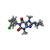  Mass: 424.281 Da / Num. of mol.: 12 / Source method: obtained synthetically / Formula: C18H19Cl2N5O3 Mass: 424.281 Da / Num. of mol.: 12 / Source method: obtained synthetically / Formula: C18H19Cl2N5O3#3: Chemical | ChemComp-CL /   Mass: 35.453 Da / Num. of mol.: 12 / Source method: obtained synthetically / Formula: Cl Mass: 35.453 Da / Num. of mol.: 12 / Source method: obtained synthetically / Formula: Cl#4: Water | ChemComp-HOH / |  Mass: 18.015 Da / Num. of mol.: 1536 / Source method: isolated from a natural source / Formula: H2O Mass: 18.015 Da / Num. of mol.: 1536 / Source method: isolated from a natural source / Formula: H2OSequence details | PROTEIN EXPRESSED IN FUSION WITH A N-TERMINAL 9 AMINO ACID PEPTIDE CONTAINING A SIX HISTIDINE ...PROTEIN EXPRESSED IN FUSION WITH A N-TERMINAL 9 AMINO ACID PEPTIDE CONTAINING | |
|---|
-Experimental details
-Experiment
| Experiment | Method: X-RAY DIFFRACTION / Number of used crystals: 1 |
|---|
- Sample preparation
| Crystal | Density Matthews: 2.43 Å3/Da / Density % sol: 49 % / Description: NONE |
|---|---|
| Crystal grow | Temperature: 294 K / Method: vapor diffusion, hanging drop / pH: 5 Details: EQUAL VOLUMES OF MOTHER LIQUOR (0.1 M SODIUM ACETATE, PH 5, 0.75 M 1,6-HEXANEDIOL) AND PROTEIN SOLUTION (10 G/L IN 0.020 M TRIS-HCL. PH 7.5, 0.15 M SODIUM CHLORIDE, 0.005 M MAGNESIUM ...Details: EQUAL VOLUMES OF MOTHER LIQUOR (0.1 M SODIUM ACETATE, PH 5, 0.75 M 1,6-HEXANEDIOL) AND PROTEIN SOLUTION (10 G/L IN 0.020 M TRIS-HCL. PH 7.5, 0.15 M SODIUM CHLORIDE, 0.005 M MAGNESIUM CHLORIDE, 2%(V/V) DMSO, 0.0002 M INHIBITOR), VAPOR DIFFUSION, HANGING-DROP, TEMPERATURE 294K. |
-Data collection
| Diffraction | Mean temperature: 100 K |
|---|---|
| Diffraction source | Source: SYNCHROTRON / Site: ESRF  / Beamline: ID14-2 / Wavelength: 0.933 / Beamline: ID14-2 / Wavelength: 0.933 |
| Detector | Type: ADSC CCD / Detector: CCD / Date: May 12, 2007 |
| Radiation | Protocol: SINGLE WAVELENGTH / Monochromatic (M) / Laue (L): M / Scattering type: x-ray |
| Radiation wavelength | Wavelength: 0.933 Å / Relative weight: 1 |
| Reflection | Resolution: 2.55→29.9 Å / Num. obs: 200628 / % possible obs: 97.6 % / Observed criterion σ(I): 0 / Redundancy: 4.2 % / Biso Wilson estimate: 45 Å2 / Rmerge(I) obs: 0.09 / Net I/σ(I): 12.3 |
| Reflection shell | Resolution: 2.55→2.65 Å / Redundancy: 4.2 % / Rmerge(I) obs: 0.45 / Mean I/σ(I) obs: 3.8 / % possible all: 95.6 |
- Processing
| Software |
| ||||||||||||||||||||||||||||||||||||||||||||||||||||||||||||||||||||||||||||||||||||||||||||||||||||||||||||||||||||||||||||||||||||||||||||||||||||||||||||||||||||||||||||||||||||||
|---|---|---|---|---|---|---|---|---|---|---|---|---|---|---|---|---|---|---|---|---|---|---|---|---|---|---|---|---|---|---|---|---|---|---|---|---|---|---|---|---|---|---|---|---|---|---|---|---|---|---|---|---|---|---|---|---|---|---|---|---|---|---|---|---|---|---|---|---|---|---|---|---|---|---|---|---|---|---|---|---|---|---|---|---|---|---|---|---|---|---|---|---|---|---|---|---|---|---|---|---|---|---|---|---|---|---|---|---|---|---|---|---|---|---|---|---|---|---|---|---|---|---|---|---|---|---|---|---|---|---|---|---|---|---|---|---|---|---|---|---|---|---|---|---|---|---|---|---|---|---|---|---|---|---|---|---|---|---|---|---|---|---|---|---|---|---|---|---|---|---|---|---|---|---|---|---|---|---|---|---|---|---|---|
| Refinement | Method to determine structure: MOLECULAR REPLACEMENT Starting model: PDB ENTRY 1HTO Resolution: 2.55→29.95 Å / Cor.coef. Fo:Fc: 0.924 / Cor.coef. Fo:Fc free: 0.915 / SU B: 10.94 / SU ML: 0.233 / Cross valid method: THROUGHOUT / ESU R: 1.195 / ESU R Free: 0.31 / Stereochemistry target values: MAXIMUM LIKELIHOOD Details: HYDROGENS HAVE BEEN ADDED IN THE RIDING POSITIONS. AMINO ACIDS 63 TO 66 AND 405 TO 412 WERE OMITTED FROM THE STRUCTURE.
| ||||||||||||||||||||||||||||||||||||||||||||||||||||||||||||||||||||||||||||||||||||||||||||||||||||||||||||||||||||||||||||||||||||||||||||||||||||||||||||||||||||||||||||||||||||||
| Solvent computation | Ion probe radii: 0.8 Å / Shrinkage radii: 0.8 Å / VDW probe radii: 1.2 Å / Solvent model: MASK | ||||||||||||||||||||||||||||||||||||||||||||||||||||||||||||||||||||||||||||||||||||||||||||||||||||||||||||||||||||||||||||||||||||||||||||||||||||||||||||||||||||||||||||||||||||||
| Displacement parameters | Biso mean: 41.987 Å2
| ||||||||||||||||||||||||||||||||||||||||||||||||||||||||||||||||||||||||||||||||||||||||||||||||||||||||||||||||||||||||||||||||||||||||||||||||||||||||||||||||||||||||||||||||||||||
| Refinement step | Cycle: LAST / Resolution: 2.55→29.95 Å
| ||||||||||||||||||||||||||||||||||||||||||||||||||||||||||||||||||||||||||||||||||||||||||||||||||||||||||||||||||||||||||||||||||||||||||||||||||||||||||||||||||||||||||||||||||||||
| Refine LS restraints |
|