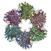
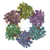
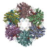
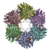
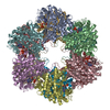
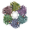
A: PROTEIN (GLUTAMINE SYNTHETASE) B: PROTEIN (GLUTAMINE SYNTHETASE) C: PROTEIN (GLUTAMINE SYNTHETASE) D: PROTEIN (GLUTAMINE SYNTHETASE) E: PROTEIN (GLUTAMINE SYNTHETASE) F: PROTEIN (GLUTAMINE SYNTHETASE) G: PROTEIN (GLUTAMINE SYNTHETASE) H: PROTEIN (GLUTAMINE SYNTHETASE) I: PROTEIN (GLUTAMINE SYNTHETASE) J: PROTEIN (GLUTAMINE SYNTHETASE) K: PROTEIN (GLUTAMINE SYNTHETASE) L: PROTEIN (GLUTAMINE SYNTHETASE) hetero molecules
Type: RIGAKU RAXIS IV / Detector: IMAGE PLATE / Date: Mar 31, 1997
Radiation
Protocol: SINGLE WAVELENGTH / Monochromatic (M) / Laue (L): M / Scattering type: x-ray
Radiation wavelength
Wavelength: 1.5418 Å / Relative weight: 1
Reflection
Resolution: 2.67→32 Å / Num. obs: 268069 / % possible obs: 82 % / Observed criterion σ(I): 0 / Redundancy: 2 % / Biso Wilson estimate: 44 Å2 / Rmerge(I) obs: 0.13 / Net I/σ(I): 7.8
Reflection shell
Resolution: 2.67→2.8 Å / Redundancy: 1.8 % / Rmerge(I) obs: 0.43 / % possible all: 55
Reflection
*PLUS
Highest resolution: 2.67 Å / Lowest resolution: 32 Å / Num. obs: 130547
-
Processing
Software
Name
Version
Classification
X-PLOR
modelbuilding
X-PLOR
3.843
refinement
DENZO
datareduction
SCALEPACK
datascaling
X-PLOR
phasing
Refinement
Method to determine structure: MOLECULAR REPLACEMEN / Resolution: 2.67→32 Å / σ(F): 0 Details: THE MODEL WAS REFINED USING STRICT 12-FOLD NCS-AVERAGING, I.E. CONSTRAINTS. THE 2- METHYL-2,4-PENTANEDIOL (MPD) MOLECULE WAS RESTRAINED TO MATCH PD CODE 3AL1. A BULK SOLVENT CORRECTION WAS ...Details: THE MODEL WAS REFINED USING STRICT 12-FOLD NCS-AVERAGING, I.E. CONSTRAINTS. THE 2- METHYL-2,4-PENTANEDIOL (MPD) MOLECULE WAS RESTRAINED TO MATCH PD CODE 3AL1. A BULK SOLVENT CORRECTION WAS APPLIED TO THE DATA SET. METHOD USED: J.-S. JIANG AND A.T. BRUNGER, J. MOL. BIOL. 243, 100-115 (1994).
Rfactor
Num. reflection
% reflection
Selection details
Rfree
0.263
6529
-
RANDOM
Rwork
0.232
-
-
-
obs
0.232
130596
82 %
-
Displacement parameters
Biso mean: 46.43 Å2
Refine analyze
Free
Obs
Luzzati coordinate error
0.42 Å
0.37 Å
Luzzati d res low
-
7 Å
Refinement step
Cycle: LAST / Resolution: 2.67→32 Å
Protein
Nucleic acid
Ligand
Solvent
Total
Num. atoms
43644
0
372
1548
45564
Refine LS restraints
Refine-ID
Type
Dev ideal
X-RAY DIFFRACTION
x_bond_d
0.01
X-RAY DIFFRACTION
x_bond_d_na
X-RAY DIFFRACTION
x_bond_d_prot
X-RAY DIFFRACTION
x_angle_d
X-RAY DIFFRACTION
x_angle_d_na
X-RAY DIFFRACTION
x_angle_d_prot
X-RAY DIFFRACTION
x_angle_deg
1.79
X-RAY DIFFRACTION
x_angle_deg_na
X-RAY DIFFRACTION
x_angle_deg_prot
X-RAY DIFFRACTION
x_dihedral_angle_d
25.07
X-RAY DIFFRACTION
x_dihedral_angle_d_na
X-RAY DIFFRACTION
x_dihedral_angle_d_prot
X-RAY DIFFRACTION
x_improper_angle_d
1.81
X-RAY DIFFRACTION
x_improper_angle_d_na
X-RAY DIFFRACTION
x_improper_angle_d_prot
X-RAY DIFFRACTION
x_mcbond_it
X-RAY DIFFRACTION
x_mcangle_it
X-RAY DIFFRACTION
x_scbond_it
X-RAY DIFFRACTION
x_scangle_it
Refine LS restraints NCS
NCS model details: CONSTRAINED
LS refinement shell
Resolution: 2.67→2.79 Å / Total num. of bins used: 8
In the structure databanks used in Yorodumi, some data are registered as the other names, "COVID-19 virus" and "2019-nCoV". Here are the details of the virus and the list of structure data.
Jan 31, 2019. EMDB accession codes are about to change! (news from PDBe EMDB page)
EMDB accession codes are about to change! (news from PDBe EMDB page)
The allocation of 4 digits for EMDB accession codes will soon come to an end. Whilst these codes will remain in use, new EMDB accession codes will include an additional digit and will expand incrementally as the available range of codes is exhausted. The current 4-digit format prefixed with “EMD-” (i.e. EMD-XXXX) will advance to a 5-digit format (i.e. EMD-XXXXX), and so on. It is currently estimated that the 4-digit codes will be depleted around Spring 2019, at which point the 5-digit format will come into force.
The EM Navigator/Yorodumi systems omit the EMD- prefix.
Related info.:Q: What is EMD? / ID/Accession-code notation in Yorodumi/EM Navigator
Yorodumi is a browser for structure data from EMDB, PDB, SASBDB, etc.
This page is also the successor to EM Navigator detail page, and also detail information page/front-end page for Omokage search.
The word "yorodu" (or yorozu) is an old Japanese word meaning "ten thousand". "mi" (miru) is to see.
Related info.:EMDB / PDB / SASBDB / Comparison of 3 databanks / Yorodumi Search / Aug 31, 2016. New EM Navigator & Yorodumi / Yorodumi Papers / Jmol/JSmol / Function and homology information / Changes in new EM Navigator and Yorodumi
 Movie
Movie Controller

 Yorodumi
 Open data
 Basic information
 Components
 Keywords
 Function and homology information
 Salmonella typhimurium (bacteria)
 X-RAY DIFFRACTION / MOLECULAR REPLACEMEN / Resolution: 2.67 Å
  Authors
 Citation
 Structure visualization
 Downloads & links
 Other downloads
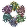


 PDBj
PDBj
 Assembly
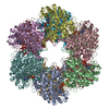


 Mass: 54.938 Da / Num. of mol.: 24 / Source method: obtained synthetically / Formula: Mn
 Mass: 427.201 Da / Num. of mol.: 12 / Source method: obtained synthetically / Formula: C10H15N5O10P2 / Comment: ADP, energy-carrying molecule*YM
 Mass: 204.383 Da / Num. of mol.: 24 / Source method: obtained synthetically / Formula: Tl
 Mass: 118.174 Da / Num. of mol.: 12 / Source method: obtained synthetically / Formula: C6H14O2 / Comment: precipitant*YM
 Sample preparation
 Processing