 Movie
Movie Controller

[English] 日本語
 Yorodumi
- PDB-2gls: REFINED ATOMIC MODEL OF GLUTAMINE SYNTHETASE AT 3.5 ANGSTROMS RES... -
+ Open data

- Basic information
| Entry | Database: PDB / ID: 2gls | |||||||||
|---|---|---|---|---|---|---|---|---|---|---|
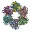
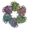
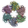
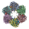
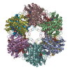
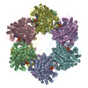
| Title | REFINED ATOMIC MODEL OF GLUTAMINE SYNTHETASE AT 3.5 ANGSTROMS RESOLUTION | |||||||||
 Components Components | GLUTAMINE SYNTHETASE | |||||||||
 Keywords Keywords | LIGASE(AMIDE SYNTHETASE) | |||||||||
| Function / homology |  Function and homology information Function and homology informationnitrogen utilization / glutamine synthetase / : / glutamine synthetase activity / protein homooligomerization / manganese ion binding / ATP binding / membrane / cytoplasm Similarity search - Function | |||||||||
| Biological species |  Salmonella typhimurium (bacteria) Salmonella typhimurium (bacteria) | |||||||||
| Method |  X-RAY DIFFRACTION / Resolution: 3.5 Å X-RAY DIFFRACTION / Resolution: 3.5 Å | |||||||||
 Authors Authors | Eisenberg, D. / Almassy, R.J. / Yamashita, M.M. | |||||||||
 Citation Citation | Journal: J.Biol.Chem. / Year: 1989 Title: Refined atomic model of glutamine synthetase at 3.5 A resolution. Authors: Yamashita, M.M. / Almassy, R.J. / Janson, C.A. / Cascio, D. / Eisenberg, D. #1: Journal: Nature / Year: 1986Title: Novel Subunit-Subunit Interactions in the Structure of Glutamine Synthetase Authors: Almassy, R.J. / Janson, C.A. / Hamlin, R. / Xuong, N.-H. / Eisenberg, D. #2: Journal: Gene / Year: 1986Title: Sequence of Glutamine Synthetase from Salmonella Typhimurium and Implications for the Protein Structure Authors: Janson, C.A. / Kayne, P.S. / Almassy, R.J. / Grunstein, M. / Eisenberg, D. | |||||||||
| History |
|
- Structure visualization
| Structure viewer | Molecule: MolmilJmol/JSmol |
|---|
- Downloads & links
-Download
| PDBx/mmCIF format | 2gls.cif.gz | 972.5 KB | Display | PDBx/mmCIF format |
|---|---|---|---|---|
| PDB format | pdb2gls.ent.gz | 775.4 KB | Display | PDB format |
| PDBx/mmJSON format | 2gls.json.gz | Tree view | PDBx/mmJSON format | |
| Others |  Other downloads Other downloads |
-Validation report
| Arichive directory | https://data.pdbj.org/pub/pdb/validation_reports/gl/2glsftp://data.pdbj.org/pub/pdb/validation_reports/gl/2gls | HTTPS FTP |
|---|
-Related structure data
| Similar structure data |
|---|


-Links
 PDBj
PDBj
- Assembly
| Deposited unit | 
| ||||||||
|---|---|---|---|---|---|---|---|---|---|
| 1 |
| ||||||||
| Unit cell |
|
-Components
| #1: Protein | Mass: 51875.609 Da / Num. of mol.: 12 Source method: isolated from a genetically manipulated source Source: (gene. exp.) Salmonella typhimurium (bacteria) / References: UniProt: P0A1P6, glutamine synthetase#2: Chemical | ChemComp-MN /   Mass: 54.938 Da / Num. of mol.: 24 / Source method: obtained synthetically / Formula: Mn Mass: 54.938 Da / Num. of mol.: 24 / Source method: obtained synthetically / Formula: Mn#3: Water | ChemComp-HOH / |  Mass: 18.015 Da / Num. of mol.: 36 / Source method: isolated from a natural source / Formula: H2O Mass: 18.015 Da / Num. of mol.: 36 / Source method: isolated from a natural source / Formula: H2O |
|---|
-Experimental details
-Experiment
| Experiment | Method: X-RAY DIFFRACTION |
|---|
- Sample preparation
| Crystal | Density Matthews: 2.48 Å3/Da / Density % sol: 50.42 % | ||||||||||||||||||||||||||||||||||||
|---|---|---|---|---|---|---|---|---|---|---|---|---|---|---|---|---|---|---|---|---|---|---|---|---|---|---|---|---|---|---|---|---|---|---|---|---|---|
| Crystal grow | *PLUS Temperature: 21 ℃ / pH: 7.1 / Method: vapor diffusion, hanging drop / Details: took from Jonson et al.,(1984) from original paper | ||||||||||||||||||||||||||||||||||||
| Components of the solutions | *PLUS
|
-Data collection
| Radiation | Scattering type: x-ray |
|---|---|
| Radiation wavelength | Relative weight: 1 |
| Reflection | *PLUS Highest resolution: 3.5 Å / Num. all: 76889 / Num. obs: 65223 / Rmerge F obs: 0.055 |
- Processing
| Software | Name: PROLSQ / Classification: refinement | ||||||||||||||||||||||||||||||||||||||||||||||||||||||||||||||||||||||||||||||||||||
|---|---|---|---|---|---|---|---|---|---|---|---|---|---|---|---|---|---|---|---|---|---|---|---|---|---|---|---|---|---|---|---|---|---|---|---|---|---|---|---|---|---|---|---|---|---|---|---|---|---|---|---|---|---|---|---|---|---|---|---|---|---|---|---|---|---|---|---|---|---|---|---|---|---|---|---|---|---|---|---|---|---|---|---|---|---|
| Refinement | Rfactor obs: 0.258 / Highest resolution: 3.5 Å / σ(F): 2 | ||||||||||||||||||||||||||||||||||||||||||||||||||||||||||||||||||||||||||||||||||||
| Refinement step | Cycle: LAST / Highest resolution: 3.5 Å
| ||||||||||||||||||||||||||||||||||||||||||||||||||||||||||||||||||||||||||||||||||||
| Refine LS restraints |
| ||||||||||||||||||||||||||||||||||||||||||||||||||||||||||||||||||||||||||||||||||||
| Software | *PLUS Name: PROLSQ / Classification: refinement | ||||||||||||||||||||||||||||||||||||||||||||||||||||||||||||||||||||||||||||||||||||
| Refinement | *PLUS Lowest resolution: 10 Å / Num. reflection obs: 65223 / Rfactor obs: 0.258 | ||||||||||||||||||||||||||||||||||||||||||||||||||||||||||||||||||||||||||||||||||||
| Solvent computation | *PLUS | ||||||||||||||||||||||||||||||||||||||||||||||||||||||||||||||||||||||||||||||||||||
| Displacement parameters | *PLUS Biso mean: 25 Å2 |
