 Movie
Movie Controller

[English] 日本語
 Yorodumi
- PDB-1htq: Multicopy crystallographic structure of a relaxed glutamine synth... -
+ Open data

- Basic information
| Entry | Database: PDB / ID: 1htq | ||||||
|---|---|---|---|---|---|---|---|
| Title | Multicopy crystallographic structure of a relaxed glutamine synthetase from Mycobacterium tuberculosis | ||||||
 Components Components | glutamine synthetase | ||||||
 Keywords Keywords | LIGASE / glutamine synthetase / Mycobacterium tuberculosis / multicopy refinement / Structural Genomics / PSI / Protein Structure Initiative / TB Structural Genomics Consortium / TBSGC | ||||||
| Function / homology |  Function and homology information Function and homology informationnitrogen utilization / positive regulation of plasminogen activation / glutamine synthetase / : / glutamine synthetase activity / zymogen binding / cobalt ion binding / fibronectin binding / peptidoglycan-based cell wall / ADP binding ...nitrogen utilization / positive regulation of plasminogen activation / glutamine synthetase / : / glutamine synthetase activity / zymogen binding / cobalt ion binding / fibronectin binding / peptidoglycan-based cell wall / ADP binding / protein homooligomerization / GDP binding / manganese ion binding / GTP binding / magnesium ion binding / extracellular region / ATP binding / membrane / metal ion binding / plasma membrane / cytoplasm / cytosol Similarity search - Function | ||||||
| Biological species |   Mycobacterium tuberculosis (bacteria) Mycobacterium tuberculosis (bacteria) | ||||||
| Method |  X-RAY DIFFRACTION / SYNCHROTRON / Multicopy Refinement / Resolution: 2.4 Å X-RAY DIFFRACTION / SYNCHROTRON / Multicopy Refinement / Resolution: 2.4 Å | ||||||
 Authors Authors | Gill, H.S. / Pfluegl, G.M. / Eisenberg, D. / TB Structural Genomics Consortium (TBSGC) | ||||||
 Citation Citation | Journal: Biochemistry / Year: 2002 Title: Multicopy crystallographic refinement of a relaxed glutamine synthetase from Mycobacterium tuberculosis highlights flexible loops in the enzymatic mechanism and its regulation. Authors: Gill, H.S. / Pfluegl, G.M. / Eisenberg, D. | ||||||
| History |
|
- Structure visualization
| Structure viewer | Molecule: MolmilJmol/JSmol |
|---|
- Downloads & links
-Download
| PDBx/mmCIF format | 1htq.cif.gz | 21.7 MB | Display | PDBx/mmCIF format |
|---|---|---|---|---|
| PDB format | pdb1htq.ent.gz | 18.6 MB | Display | PDB format |
| PDBx/mmJSON format | 1htq.json.gz | Tree view | PDBx/mmJSON format | |
| Others |  Other downloads Other downloads |
-Validation report
| Arichive directory | https://data.pdbj.org/pub/pdb/validation_reports/ht/1htqftp://data.pdbj.org/pub/pdb/validation_reports/ht/1htq | HTTPS FTP |
|---|
-Related structure data
| Related structure data | 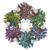 1htoSC S: Starting model for refinement C: citing same article ( |
|---|---|
| Similar structure data | |
| Other databases |


-Links
 PDBj
PDBj


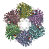
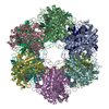
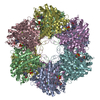
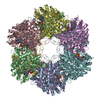
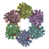
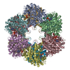
- Assembly
| Deposited unit | 
| ||||||||
|---|---|---|---|---|---|---|---|---|---|
| 1 | 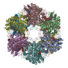
| ||||||||
| 2 | 
| ||||||||
| Unit cell |
| ||||||||
| Number of models | 10 | ||||||||
| Details | The biological complex is a dodecamer. |
-Components
| #1: Protein | Mass: 53496.531 Da / Num. of mol.: 24 Source method: isolated from a genetically manipulated source Source: (gene. exp.) Mycobacterium tuberculosis (bacteria) / Plasmid: pTrcHisB / Production host: References: UniProt: Q10377, UniProt: P9WN39*PLUS, glutamine synthetase #2: Chemical | ChemComp-MN /   Mass: 54.938 Da / Num. of mol.: 24 / Source method: obtained synthetically / Formula: Mn Mass: 54.938 Da / Num. of mol.: 24 / Source method: obtained synthetically / Formula: Mn#3: Chemical | ChemComp-AMP /   Mass: 347.221 Da / Num. of mol.: 24 / Source method: obtained synthetically / Formula: C10H14N5O7P / Comment: AMP*YM Mass: 347.221 Da / Num. of mol.: 24 / Source method: obtained synthetically / Formula: C10H14N5O7P / Comment: AMP*YM#4: Chemical | ChemComp-CIT /   Mass: 192.124 Da / Num. of mol.: 24 / Source method: obtained synthetically / Formula: C6H8O7 Mass: 192.124 Da / Num. of mol.: 24 / Source method: obtained synthetically / Formula: C6H8O7#5: Water | ChemComp-HOH / |  Mass: 18.015 Da / Num. of mol.: 6312 / Source method: isolated from a natural source / Formula: H2O Mass: 18.015 Da / Num. of mol.: 6312 / Source method: isolated from a natural source / Formula: H2O |
|---|
-Experimental details
-Experiment
| Experiment | Method: X-RAY DIFFRACTION / Number of used crystals: 1 |
|---|
- Sample preparation
| Crystal | Density Matthews: 2.86 Å3/Da / Density % sol: 56.98 % |
|---|---|
| Crystal grow | Temperature: 298 K / Method: vapor diffusion, hanging drop / pH: 5.5 Details: PEG 4000, sodium citrate, sodium chloride, manganese chloride, imidazole buffer , pH 5.5, VAPOR DIFFUSION, HANGING DROP, temperature 298K |
-Data collection
| Diffraction | Mean temperature: 100 K |
|---|---|
| Diffraction source | Source: SYNCHROTRON / Site: NSLS  / Beamline: X12B / Wavelength: 0.978 Å / Beamline: X12B / Wavelength: 0.978 Å |
| Detector | Type: ADSC QUANTUM 4 / Detector: CCD / Date: Jul 1, 1997 |
| Radiation | Protocol: SINGLE WAVELENGTH / Monochromatic (M) / Laue (L): M / Scattering type: x-ray |
| Radiation wavelength | Wavelength: 0.978 Å / Relative weight: 1 |
| Reflection | Resolution: 2.4→20 Å / Num. all: 566370 / Num. obs: 566370 / % possible obs: 99.6 % / Observed criterion σ(F): 0 / Observed criterion σ(I): 0 / Redundancy: 4.6 % / Biso Wilson estimate: 33.1 Å2 / Rmerge(I) obs: 0.075 / Net I/σ(I): 4.5 |
| Reflection shell | Resolution: 2.4→2.53 Å / Redundancy: 3.6 % / Rmerge(I) obs: 0.075 / Num. unique all: 80442 / % possible all: 98.8 |
- Processing
| Software |
| |||||||||||||||||||||||||
|---|---|---|---|---|---|---|---|---|---|---|---|---|---|---|---|---|---|---|---|---|---|---|---|---|---|---|
| Refinement | Method to determine structure: Multicopy Refinement Starting model: 1HTO Resolution: 2.4→20 Å / σ(F): 0 / σ(I): 0 / Stereochemistry target values: Engh & Huber Details: THE AVERAGED-SUBUNIT (MONOMERIC) MODEL OF THE PDB ENTRY 1HTO WAS FURTHER REFINED HERE BY DUPLICATING 10 COPIES OF THE MONOMERIC MODEL WITH 24-FOLD STRICT NCS-AVERAGING CONSTRAINTS IMPOSED ON ...Details: THE AVERAGED-SUBUNIT (MONOMERIC) MODEL OF THE PDB ENTRY 1HTO WAS FURTHER REFINED HERE BY DUPLICATING 10 COPIES OF THE MONOMERIC MODEL WITH 24-FOLD STRICT NCS-AVERAGING CONSTRAINTS IMPOSED ON EACH MODEL. THE ENTIRE ENSEMBLE WAS THEN SIMULTANEOUSLY REFINED AGAINST THE DATA BY SIMULATED ANNEALING PROTOCOLS WITH A FIXED OVERALL B-FACTOR AND 0.1 PARTIAL OCCUPANCIES ASSIGNED TO EACH OF THE TEN MODELS, USING PROGRAM XPLOR 3.843. CHEMICALLY EACH COPY OR CHAIN DOES NOT SEE ONE ANOTHER DURING REFINEMENT, BUT EACH ADDS TOGETHER TO CALCULATE THE TOTAL STRUCTURE FACTOR. POSITIONAL AND B-FACTOR REFINEMENT WERE OPTIONALLY EMPLOYEED TO EACH OF THE 10 MODELS THEREAFTER. LIGANDS AND WATERS WERE ALSO OPTIONALLY FIXED DURING THE REFINEMENT. THIS REPRESENTATIVE ENTRY EMPLOYEED POSITIONAL AND B-FACTOR REFINEMENT AFTER SIMULATED ANNEALING WITH ONLY THE LIGANDS FIXED DURING REFINEMENT. ALL RESULTS INDICATED A DROP IN BOTH THE R-FACTOR AND R-FREE FROM THE 1HT0 ENTRY, AND APPEAR STRUCTURALLY SIMILAR. THE FULL ASYMMETRIC UNIT WAS SUBSEQUENTLY GENERATED FOR EACH OF THE 10 COPIES BY APPLYING THE 24-FOLD NCS-SYMMETRY AND IN TOTAL CONTAINS APPROXIMATELY 1 MILLION ATOMS. THE MULTICOPY MODEL HIGHLIGHTS MOVEMENTS OF CATALYTIC AND REGULATORY LOOPS IN GLUTAMINE SYNTHESIS.
| |||||||||||||||||||||||||
| Refinement step | Cycle: LAST / Resolution: 2.4→20 Å
| |||||||||||||||||||||||||
| Refine LS restraints |
|
