 Movie
Movie Controller

[English] 日本語
 Yorodumi
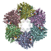
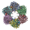
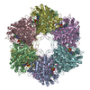
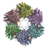
- PDB-1f52: CRYSTAL STRUCTURE OF GLUTAMINE SYNTHETASE FROM SALMONELLA TYPHIMU... -
+ Open data

- Basic information
| Entry | Database: PDB / ID: 1f52 | ||||||
|---|---|---|---|---|---|---|---|
| Title | CRYSTAL STRUCTURE OF GLUTAMINE SYNTHETASE FROM SALMONELLA TYPHIMURIUM CO-CRYSTALLIZED WITH ADP | ||||||
 Components Components | GLUTAMINE SYNTHETASE | ||||||
 Keywords Keywords | LIGASE / glutamine synthetase / ADP / MPD | ||||||
| Function / homology |  Function and homology information Function and homology informationnitrogen utilization / glutamine synthetase / : / glutamine synthetase activity / protein homooligomerization / manganese ion binding / ATP binding / membrane / cytoplasm Similarity search - Function | ||||||
| Biological species |  Salmonella typhimurium (bacteria) Salmonella typhimurium (bacteria) | ||||||
| Method |  X-RAY DIFFRACTION / SYNCHROTRON / MOLECULAR REPLACEMENT / Resolution: 2.49 Å X-RAY DIFFRACTION / SYNCHROTRON / MOLECULAR REPLACEMENT / Resolution: 2.49 Å | ||||||
 Authors Authors | Gill, H.S. / Pfluegl, G.M.U. / Eisenberg, D. | ||||||
 Citation Citation | Journal: Biochemistry / Year: 2001 Title: The crystal structure of phosphinothricin in the active site of glutamine synthetase illuminates the mechanism of enzymatic inhibition. Authors: Gill, H.S. / Eisenberg, D. | ||||||
| History |
|
- Structure visualization
| Structure viewer | Molecule: MolmilJmol/JSmol |
|---|
- Downloads & links
-Download
| PDBx/mmCIF format | 1f52.cif.gz | 1.1 MB | Display | PDBx/mmCIF format |
|---|---|---|---|---|
| PDB format | pdb1f52.ent.gz | 950.6 KB | Display | PDB format |
| PDBx/mmJSON format | 1f52.json.gz | Tree view | PDBx/mmJSON format | |
| Others |  Other downloads Other downloads |
-Validation report
| Arichive directory | https://data.pdbj.org/pub/pdb/validation_reports/f5/1f52ftp://data.pdbj.org/pub/pdb/validation_reports/f5/1f52 | HTTPS FTP |
|---|
-Related structure data


-Links
 PDBj
PDBj
- Assembly
| Deposited unit | 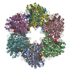
| ||||||||
|---|---|---|---|---|---|---|---|---|---|
| 1 |
| ||||||||
| Unit cell |
| ||||||||
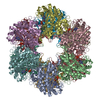
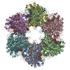
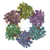
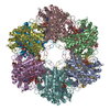
| Details | The biological complex is a dodecamer. |
-Components
| #1: Protein | Mass: 51744.418 Da / Num. of mol.: 12 Source method: isolated from a genetically manipulated source Source: (gene. exp.) Salmonella typhimurium (bacteria) / Description: BACTERIA / Production host: #2: Chemical | ChemComp-MN /   Mass: 54.938 Da / Num. of mol.: 24 / Source method: obtained synthetically / Formula: Mn Mass: 54.938 Da / Num. of mol.: 24 / Source method: obtained synthetically / Formula: Mn#3: Chemical | ChemComp-ADP /   Mass: 427.201 Da / Num. of mol.: 12 / Source method: obtained synthetically / Formula: C10H15N5O10P2 / Comment: ADP, energy-carrying molecule*YM Mass: 427.201 Da / Num. of mol.: 12 / Source method: obtained synthetically / Formula: C10H15N5O10P2 / Comment: ADP, energy-carrying molecule*YM#4: Chemical | ChemComp-MPD / (   Mass: 118.174 Da / Num. of mol.: 24 / Source method: obtained synthetically / Formula: C6H14O2 / Comment: precipitant*YM Mass: 118.174 Da / Num. of mol.: 24 / Source method: obtained synthetically / Formula: C6H14O2 / Comment: precipitant*YM#5: Water | ChemComp-HOH / |  Mass: 18.015 Da / Num. of mol.: 3504 / Source method: isolated from a natural source / Formula: H2O Mass: 18.015 Da / Num. of mol.: 3504 / Source method: isolated from a natural source / Formula: H2O |
|---|
-Experimental details
-Experiment
| Experiment | Method: X-RAY DIFFRACTION / Number of used crystals: 1 |
|---|
- Sample preparation
| Crystal | Density Matthews: 2.36 Å3/Da / Density % sol: 47.8 % | ||||||||||||||||||||||||||||||
|---|---|---|---|---|---|---|---|---|---|---|---|---|---|---|---|---|---|---|---|---|---|---|---|---|---|---|---|---|---|---|---|
| Crystal grow | Temperature: 298 K / Method: vapor diffusion, hanging drop / pH: 7.5 Details: ADP, MPD, spermine, manganese chloride, imidazole buffer, pH 7.5, VAPOR DIFFUSION, HANGING DROP, temperature 298K | ||||||||||||||||||||||||||||||
| Crystal grow | *PLUS pH: 7 | ||||||||||||||||||||||||||||||
| Components of the solutions | *PLUS
|
-Data collection
| Diffraction | Mean temperature: 100 K |
|---|---|
| Diffraction source | Source: SYNCHROTRON / Site: NSLS  / Beamline: X12C / Wavelength: 1.1 / Beamline: X12C / Wavelength: 1.1 |
| Detector | Type: MARRESEARCH / Detector: IMAGE PLATE / Date: Apr 24, 1995 |
| Radiation | Protocol: SINGLE WAVELENGTH / Monochromatic (M) / Laue (L): M / Scattering type: x-ray |
| Radiation wavelength | Wavelength: 1.1 Å / Relative weight: 1 |
| Reflection | Resolution: 2.49→34.9 Å / Num. all: 196561 / Num. obs: 196561 / % possible obs: 98.2 % / Observed criterion σ(F): 0 / Observed criterion σ(I): 0 / Redundancy: 10 % / Biso Wilson estimate: 56.6 Å2 / Rmerge(I) obs: 0.082 / Net I/σ(I): 32.6 |
| Reflection shell | Resolution: 2.49→2.53 Å / Redundancy: 2.3 % / Rmerge(I) obs: 0.133 / Num. unique all: 8787 / % possible all: 87.8 |
| Reflection | *PLUS |
- Processing
| Software |
| |||||||||||||||||||||||||
|---|---|---|---|---|---|---|---|---|---|---|---|---|---|---|---|---|---|---|---|---|---|---|---|---|---|---|
| Refinement | Method to determine structure: MOLECULAR REPLACEMENT / Resolution: 2.49→34.9 Å / σ(F): 0 / σ(I): 0 / Stereochemistry target values: Engh & Huber Details: The model was refined using strict 12-fold NCS-averaging, i.e. constraints. The 2-methyl-2,4-pentanediol (MPD) molecules were restrained to match PDB code 3AL1. A bulk solvent correction was ...Details: The model was refined using strict 12-fold NCS-averaging, i.e. constraints. The 2-methyl-2,4-pentanediol (MPD) molecules were restrained to match PDB code 3AL1. A bulk solvent correction was applied to the data set. Method Used: J.-S. Jiang and A.T. Brunger, J. Mol. Biol. 243, 100-115 (1994).
| |||||||||||||||||||||||||
| Displacement parameters | Biso mean: 55.54 Å2
| |||||||||||||||||||||||||
| Refine analyze |
| |||||||||||||||||||||||||
| Refinement step | Cycle: LAST / Resolution: 2.49→34.9 Å
| |||||||||||||||||||||||||
| Refine LS restraints |
| |||||||||||||||||||||||||
| Refine LS restraints NCS | NCS model details: CONSTRAINED | |||||||||||||||||||||||||
| Xplor file | Serial no: 1 / Param file: parhcsdx.pro / Topol file: tophcsdx.pro | |||||||||||||||||||||||||
| Software | *PLUS Name: X-PLOR / Version: 3.843 / Classification: refinement | |||||||||||||||||||||||||
| Refinement | *PLUS Lowest resolution: 34.9 Å / σ(F): 0 | |||||||||||||||||||||||||
| Solvent computation | *PLUS | |||||||||||||||||||||||||
| Displacement parameters | *PLUS | |||||||||||||||||||||||||
| Refine LS restraints | *PLUS
|
