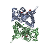
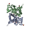
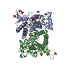
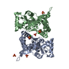
- PDB-4dld: Crystal structure of the GluK1 ligand-binding domain (S1S2) in co... -
+
Open data
ID or keywords:
Loading...
-
Basic information
Entry
Database: PDB / ID: 4dld
Title
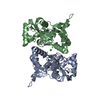
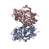
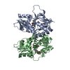
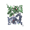
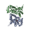
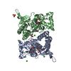
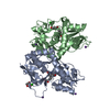
Crystal structure of the GluK1 ligand-binding domain (S1S2) in complex with the antagonist (S)-2-amino-3-(2-(2-carboxyethyl)-5-chloro-4-nitrophenyl)propionic acid at 2.0 A resolution
Components
Glutamate receptor, ionotropic kainate 1
Keywords
MEMBRANE PROTEIN / ionotropic glutamate receptor / glutamate / membrane
Function / homology
Function and homology information
negative regulation of synaptic transmission, GABAergic / L-glutamate transmembrane transporter activity / positive regulation of gamma-aminobutyric acid secretion / Activation of Na-permeable kainate receptors / Activation of Ca-permeable Kainate Receptor / kainate selective glutamate receptor complex / regulation of short-term neuronal synaptic plasticity / negative regulation of synaptic transmission, glutamatergic / glutamate binding / inhibitory postsynaptic potential ...negative regulation of synaptic transmission, GABAergic / L-glutamate transmembrane transporter activity / positive regulation of gamma-aminobutyric acid secretion / Activation of Na-permeable kainate receptors / Activation of Ca-permeable Kainate Receptor / kainate selective glutamate receptor complex / regulation of short-term neuronal synaptic plasticity / negative regulation of synaptic transmission, glutamatergic / glutamate binding / inhibitory postsynaptic potential / adult behavior / kainate selective glutamate receptor activity / behavioral response to pain / extracellularly glutamate-gated ion channel activity / modulation of excitatory postsynaptic potential / ionotropic glutamate receptor complex / membrane depolarization / glutamate-gated receptor activity / glutamate-gated calcium ion channel activity / establishment of localization in cell / ionotropic glutamate receptor signaling pathway / ligand-gated monoatomic ion channel activity involved in regulation of presynaptic membrane potential / presynaptic modulation of chemical synaptic transmission / positive regulation of synaptic transmission, GABAergic / SNARE binding / synaptic transmission, glutamatergic / transmitter-gated monoatomic ion channel activity involved in regulation of postsynaptic membrane potential / regulation of membrane potential / excitatory postsynaptic potential / regulation of synaptic plasticity / postsynaptic density membrane / modulation of chemical synaptic transmission / terminal bouton / nervous system development / presynaptic membrane / scaffold protein binding / chemical synaptic transmission / postsynaptic membrane / signaling receptor complex / postsynaptic density / neuronal cell body / synapse / dendrite / glutamatergic synapse / membrane / identical protein binding / plasma membrane Similarity search - Function
Ionotropic glutamate receptor, metazoa / Ligated ion channel L-glutamate- and glycine-binding site / Ligand-gated ion channel / Ionotropic glutamate receptor, L-glutamate and glycine-binding domain / Ligated ion channel L-glutamate- and glycine-binding site / : / Ionotropic glutamate receptor / Eukaryotic homologues of bacterial periplasmic substrate binding proteins. / Periplasmic binding protein-like II / Receptor, ligand binding region ...Ionotropic glutamate receptor, metazoa / Ligated ion channel L-glutamate- and glycine-binding site / Ligand-gated ion channel / Ionotropic glutamate receptor, L-glutamate and glycine-binding domain / Ligated ion channel L-glutamate- and glycine-binding site / : / Ionotropic glutamate receptor / Eukaryotic homologues of bacterial periplasmic substrate binding proteins. / Periplasmic binding protein-like II / Receptor, ligand binding region / Receptor family ligand binding region / D-Maltodextrin-Binding Protein; domain 2 / Periplasmic binding protein-like I / 3-Layer(aba) Sandwich / Alpha Beta Similarity search - Domain/homology
Mass: 29108.453 Da / Num. of mol.: 2 / Fragment: GluK1 Ligand binding domain (GluK1-S1S2) Source method: isolated from a genetically manipulated source Details: The protein crystallized is the extracellular ligand binding domain of GluK1. Transmembrane regions were genetically removed and replaced with a Gly-Thr linker. Source: (gene. exp.) Rattus norvegicus (Norway rat) / Gene: Grik1 / Plasmid: pET28 / Production host: Escherichia coli (E. coli) / Strain (production host): Origami 2 / References: UniProt: P22756
Mass: 18.015 Da / Num. of mol.: 501 / Source method: isolated from a natural source / Formula: H2O
-
Details
Has protein modification
Y
Sequence details
THE SEQUENCE DATABASE IS P22756-2, ISOFORM GLUR5-2. THE CRYSTALLIZED PROTEIN CONSISTS OF GLY ...THE SEQUENCE DATABASE IS P22756-2, ISOFORM GLUR5-2. THE CRYSTALLIZED PROTEIN CONSISTS OF GLY RESIDUE FOLLOWED BY RESIDUE 430-544 AND 667-805 FROM THE INTACT RECEPTOR LINKED BY A GLY-THR DIPEPTIDE. THERE IS A SEQUENCE CONFLICT AT RESIDUE 34 OF THE CRYSTALLIZED PROTEIN DUE TO DIFFERENCES IN DATABASE SEQUENCE (SEE GENBANK ACCESION NO.AAA02874).
-
Experimental details
-
Experiment
Experiment
Method: X-RAY DIFFRACTION / Number of used crystals: 1
-
Sample preparation
Crystal
Density Matthews: 2.42 Å3/Da / Density % sol: 49.26 % / Mosaicity: 0.69 °
Crystal grow
Temperature: 279 K / Method: vapor diffusion, hanging drop / pH: 6.5 Details: 17 % PEG4000, 0.1M cacodylic acid, 0.3M lithium sulfate, pH 6.5, VAPOR DIFFUSION, HANGING DROP, temperature 279K
-
Data collection
Diffraction
Mean temperature: 100 K
Diffraction source
Source: SYNCHROTRON / Site: MAX II / Beamline: I911-2 / Wavelength: 1.038 Å
Detector
Type: MAR CCD 165 mm / Detector: CCD / Date: Mar 19, 2010
Radiation
Protocol: SINGLE WAVELENGTH / Monochromatic (M) / Laue (L): M / Scattering type: x-ray
Radiation wavelength
Wavelength: 1.038 Å / Relative weight: 1
Reflection
Resolution: 2→25.877 Å / Num. all: 39813 / Num. obs: 39813 / % possible obs: 99.9 % / Redundancy: 7.9 % / Biso Wilson estimate: 21.09 Å2 / Rsym value: 0.081 / Net I/σ(I): 6
Reflection shell
Diffraction-ID: 1
Resolution (Å)
Redundancy (%)
Rmerge(I) all
Rmerge(I) obs
Mean I/σ(I) obs
Num. measured all
Num. unique all
Rpim(I) all
Rrim(I) all
Rsym value
Net I/σ(I) obs
% possible all
2-2.11
8
0.34
0.318
1.8
45372
5696
0.119
0.34
0.318
6.3
100
2.11-2.24
8
0.243
0.227
1.5
43343
5433
0.084
0.243
0.227
8.8
100
2.24-2.39
8
0.191
0.178
3.2
40681
5097
0.066
0.191
0.178
10.9
100
2.39-2.58
8
0.142
0.133
4.2
37959
4762
0.049
0.142
0.133
13.8
100
2.58-2.83
8
0.108
0.101
6.4
34909
4386
0.038
0.108
0.101
17.2
100
2.83-3.16
7.9
0.077
0.072
6.9
32043
4037
0.027
0.077
0.072
22.5
100
3.16-3.65
7.9
0.056
0.053
12.2
28133
3563
0.02
0.056
0.053
28.8
100
3.65-4.47
7.8
0.057
0.053
11
23806
3043
0.02
0.057
0.053
33.7
99.8
4.47-6.32
7.7
0.063
0.059
8.8
18517
2408
0.022
0.063
0.059
39.1
99.5
6.32-25.877
7.1
0.043
0.04
13.9
9868
1388
0.016
0.043
0.04
43.1
97
-
Phasing
Phasing
Method: molecular replacement
-
Processing
Software
Name
Version
Classification
NB
MOSFLM
datareduction
SCALA
3.3.9
datascaling
PHASER
phasing
PHENIX
1.7.1_743
refinement
PDB_EXTRACT
3.1
dataextraction
Refinement
Method to determine structure: MOLECULAR REPLACEMENT Starting model: 2PBW chain A
In the structure databanks used in Yorodumi, some data are registered as the other names, "COVID-19 virus" and "2019-nCoV". Here are the details of the virus and the list of structure data.
Jan 31, 2019. EMDB accession codes are about to change! (news from PDBe EMDB page)
EMDB accession codes are about to change! (news from PDBe EMDB page)
The allocation of 4 digits for EMDB accession codes will soon come to an end. Whilst these codes will remain in use, new EMDB accession codes will include an additional digit and will expand incrementally as the available range of codes is exhausted. The current 4-digit format prefixed with “EMD-” (i.e. EMD-XXXX) will advance to a 5-digit format (i.e. EMD-XXXXX), and so on. It is currently estimated that the 4-digit codes will be depleted around Spring 2019, at which point the 5-digit format will come into force.
The EM Navigator/Yorodumi systems omit the EMD- prefix.
Related info.:Q: What is EMD? / ID/Accession-code notation in Yorodumi/EM Navigator
Yorodumi is a browser for structure data from EMDB, PDB, SASBDB, etc.
This page is also the successor to EM Navigator detail page, and also detail information page/front-end page for Omokage search.
The word "yorodu" (or yorozu) is an old Japanese word meaning "ten thousand". "mi" (miru) is to see.
Related info.:EMDB / PDB / SASBDB / Comparison of 3 databanks / Yorodumi Search / Aug 31, 2016. New EM Navigator & Yorodumi / Yorodumi Papers / Jmol/JSmol / Function and homology information / Changes in new EM Navigator and Yorodumi
 Movie
Movie Controller

 Yorodumi
 Open data
 Basic information
 Components
 Keywords
 Function and homology information
 X-RAY DIFFRACTION /
  Authors
 Citation
 Structure visualization
 Downloads & links
 Other downloads


 PDBj
PDBj

 Assembly
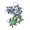


 Mass: 316.694 Da / Num. of mol.: 2 / Source method: obtained synthetically / Formula: C12H13ClN2O6
 Mass: 35.453 Da / Num. of mol.: 1 / Source method: obtained synthetically / Formula: Cl
 Mass: 96.063 Da / Num. of mol.: 1 / Source method: obtained synthetically / Formula: SO4
 Mass: 92.094 Da / Num. of mol.: 1 / Source method: obtained synthetically / Formula: C3H8O3
 Sample preparation
 / Beamline: I911-2 / Wavelength: 1.038 Å
 Processing