 Movie
Movie Controller

[English] 日本語
 Yorodumi
- PDB-3c31: Crystal structure of GluR5 ligand-binding core in complex with li... -
+ Open data

- Basic information
| Entry | Database: PDB / ID: 3c31 | ||||||
|---|---|---|---|---|---|---|---|
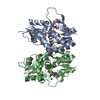
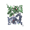
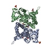
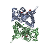
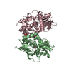
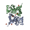
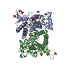
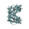
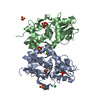
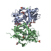
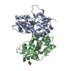
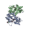
| Title | Crystal structure of GluR5 ligand-binding core in complex with lithium at 1.49 Angstrom resolution | ||||||
 Components Components | GLUTAMATE RECEPTOR, IONOTROPIC KAINATE 1 | ||||||
 Keywords Keywords | MEMBRANE PROTEIN | ||||||
| Function / homology |  Function and homology information Function and homology informationL-glutamate transmembrane transporter activity / positive regulation of gamma-aminobutyric acid secretion / Activation of Na-permeable kainate receptors / regulation of short-term neuronal synaptic plasticity / negative regulation of synaptic transmission, GABAergic / Activation of Ca-permeable Kainate Receptor / kainate selective glutamate receptor complex / negative regulation of synaptic transmission, glutamatergic / glutamate binding / inhibitory postsynaptic potential ...L-glutamate transmembrane transporter activity / positive regulation of gamma-aminobutyric acid secretion / Activation of Na-permeable kainate receptors / regulation of short-term neuronal synaptic plasticity / negative regulation of synaptic transmission, GABAergic / Activation of Ca-permeable Kainate Receptor / kainate selective glutamate receptor complex / negative regulation of synaptic transmission, glutamatergic / glutamate binding / inhibitory postsynaptic potential / adult behavior / behavioral response to pain / glutamate receptor signaling pathway / kainate selective glutamate receptor activity / extracellularly glutamate-gated ion channel activity / modulation of excitatory postsynaptic potential / ionotropic glutamate receptor complex / membrane depolarization / glutamate-gated receptor activity / glutamate-gated calcium ion channel activity / ionotropic glutamate receptor signaling pathway / positive regulation of synaptic transmission, GABAergic / ligand-gated monoatomic ion channel activity involved in regulation of presynaptic membrane potential / excitatory postsynaptic potential / SNARE binding / synaptic transmission, glutamatergic / transmitter-gated monoatomic ion channel activity involved in regulation of postsynaptic membrane potential / regulation of membrane potential / regulation of synaptic plasticity / postsynaptic density membrane / modulation of chemical synaptic transmission / terminal bouton / nervous system development / presynaptic membrane / scaffold protein binding / chemical synaptic transmission / signaling receptor complex / postsynaptic membrane / postsynaptic density / neuronal cell body / synapse / dendrite / glutamatergic synapse / membrane / identical protein binding / plasma membrane Similarity search - Function | ||||||
| Biological species |  | ||||||
| Method |  X-RAY DIFFRACTION / SYNCHROTRON / MOLECULAR REPLACEMENT / Resolution: 1.49 Å X-RAY DIFFRACTION / SYNCHROTRON / MOLECULAR REPLACEMENT / Resolution: 1.49 Å | ||||||
 Authors Authors | Mayer, M.L. | ||||||
 Citation Citation | Journal: Neuron / Year: 2008 Title: Molecular basis of kainate receptor modulation by sodium. Authors: Plested, A.J. / Vijayan, R. / Biggin, P.C. / Mayer, M.L. #1: Journal: Neuron / Year: 2007Title: Structure and mechanism of kainate receptor modulation by anions Authors: Plested, A.J. / Mayer, M.L. #2: Journal: Neuron / Year: 2005Title: Crystal structures of the GluR5 and GluR6 ligand binding cores: molecular mechanisms underlying kainate receptor selectivity. Authors: Mayer, M.L. | ||||||
| History |
|
- Structure visualization
| Structure viewer | Molecule: MolmilJmol/JSmol |
|---|
- Downloads & links
-Download
| PDBx/mmCIF format | 3c31.cif.gz | 139.4 KB | Display | PDBx/mmCIF format |
|---|---|---|---|---|
| PDB format | pdb3c31.ent.gz | 108.7 KB | Display | PDB format |
| PDBx/mmJSON format | 3c31.json.gz | Tree view | PDBx/mmJSON format | |
| Others |  Other downloads Other downloads |
-Validation report
| Arichive directory | https://data.pdbj.org/pub/pdb/validation_reports/c3/3c31ftp://data.pdbj.org/pub/pdb/validation_reports/c3/3c31 | HTTPS FTP |
|---|
-Related structure data
| Related structure data |  3c32C  3c33C  3c34C  3c35C  3c36C  1tt1S S: Starting model for refinement C: citing same article ( |
|---|---|
| Similar structure data |


-Links
 PDBj
PDBj


- Assembly
| Deposited unit | 
| ||||||||
|---|---|---|---|---|---|---|---|---|---|
| 1 |
| ||||||||
| Unit cell |
|
-Components
-Protein , 1 types, 2 molecules AB
| #1: Protein | Mass: 29253.566 Da / Num. of mol.: 2 / Fragment: Residues 446-821 Source method: isolated from a genetically manipulated source Source: (gene. exp.)  |
|---|
-Non-polymers , 6 types, 703 molecules 


| #2: Chemical |  Mass: 6.941 Da / Num. of mol.: 2 / Source method: obtained synthetically / Formula: Li Mass: 6.941 Da / Num. of mol.: 2 / Source method: obtained synthetically / Formula: Li#3: Chemical | ChemComp-CL /  Mass: 35.453 Da / Num. of mol.: 4 / Source method: obtained synthetically / Formula: Cl Mass: 35.453 Da / Num. of mol.: 4 / Source method: obtained synthetically / Formula: Cl#4: Chemical |  Mass: 213.230 Da / Num. of mol.: 2 / Source method: obtained synthetically / Formula: C10H15NO4 / Comment: neurotransmitter, agonist*YM Mass: 213.230 Da / Num. of mol.: 2 / Source method: obtained synthetically / Formula: C10H15NO4 / Comment: neurotransmitter, agonist*YM#5: Chemical | ChemComp-GOL /  Mass: 92.094 Da / Num. of mol.: 4 / Source method: obtained synthetically / Formula: C3H8O3 Mass: 92.094 Da / Num. of mol.: 4 / Source method: obtained synthetically / Formula: C3H8O3#6: Chemical | ChemComp-SO4 / |  Mass: 96.063 Da / Num. of mol.: 1 / Source method: obtained synthetically / Formula: SO4 Mass: 96.063 Da / Num. of mol.: 1 / Source method: obtained synthetically / Formula: SO4#7: Water | ChemComp-HOH / | Mass: 18.015 Da / Num. of mol.: 690 / Source method: isolated from a natural source / Formula: H2O |
|---|
-Details
| Has protein modification | Y |
|---|---|
| Sequence details | THE FIRST TWO RESIDUES OF THE SEQUENCE ARE VECTOR ENCODED. GLUR5 RESIDUES 446-559 AND 682-821 ARE ...THE FIRST TWO RESIDUES OF THE SEQUENCE ARE VECTOR ENCODED. GLUR5 RESIDUES 446-559 AND 682-821 ARE LINKED VIA GLY-THR DIPEPTIDE. |
-Experimental details
-Experiment
| Experiment | Method: X-RAY DIFFRACTION / Number of used crystals: 1 |
|---|
- Sample preparation
| Crystal | Density Matthews: 2.49 Å3/Da / Density % sol: 50.57 % |
|---|---|
| Crystal grow | Temperature: 293 K / Method: vapor diffusion, hanging drop / pH: 6 Details: 20% PEG 4000, 125 mM LiCl, 175 mM Li2(SO4), 100 mM Li cacodylate, 4 mM Kainic acid, pH 6.0, VAPOR DIFFUSION, HANGING DROP, temperature 293K |
-Data collection
| Diffraction | Mean temperature: 100 K |
|---|---|
| Diffraction source | Source: SYNCHROTRON / Site: APS  / Beamline: 22-ID / Wavelength: 1 Å / Beamline: 22-ID / Wavelength: 1 Å |
| Detector | Type: MARMOSAIC 300 mm CCD / Detector: CCD / Date: Aug 19, 2007 |
| Radiation | Monochromator: DOUBLE CRYSTAL SI-220 / Protocol: SINGLE WAVELENGTH / Monochromatic (M) / Laue (L): M / Scattering type: x-ray |
| Radiation wavelength | Wavelength: 1 Å / Relative weight: 1 |
| Reflection | Resolution: 1.49→35 Å / Num. all: 97909 / Num. obs: 97909 / % possible obs: 97.6 % / Observed criterion σ(F): 0 / Observed criterion σ(I): 0 / Redundancy: 7.3 % / Biso Wilson estimate: 16.17 Å2 / Rmerge(I) obs: 0.055 / Net I/σ(I): 13.5 |
| Reflection shell | Resolution: 1.49→1.54 Å / Redundancy: 6.5 % / Rmerge(I) obs: 0.415 / Mean I/σ(I) obs: 3.9 / % possible all: 92.2 |
- Processing
| Software |
| |||||||||||||||||||||||||||||||||||||||||||||||||||||||||||||||||||||||||||||||||||||||||||||||||||||||||||||||||||||||||||||||||||||||||||||||||||||||||||||||||||||||||||||||
|---|---|---|---|---|---|---|---|---|---|---|---|---|---|---|---|---|---|---|---|---|---|---|---|---|---|---|---|---|---|---|---|---|---|---|---|---|---|---|---|---|---|---|---|---|---|---|---|---|---|---|---|---|---|---|---|---|---|---|---|---|---|---|---|---|---|---|---|---|---|---|---|---|---|---|---|---|---|---|---|---|---|---|---|---|---|---|---|---|---|---|---|---|---|---|---|---|---|---|---|---|---|---|---|---|---|---|---|---|---|---|---|---|---|---|---|---|---|---|---|---|---|---|---|---|---|---|---|---|---|---|---|---|---|---|---|---|---|---|---|---|---|---|---|---|---|---|---|---|---|---|---|---|---|---|---|---|---|---|---|---|---|---|---|---|---|---|---|---|---|---|---|---|---|---|---|---|
| Refinement | Method to determine structure: MOLECULAR REPLACEMENT Starting model: PDB entry 1TT1 Resolution: 1.49→34.88 Å / Cor.coef. Fo:Fc: 0.967 / Cor.coef. Fo:Fc free: 0.956 / SU B: 1.867 / SU ML: 0.038 / TLS residual ADP flag: LIKELY RESIDUAL / Cross valid method: THROUGHOUT / σ(F): 0 / σ(I): 0 / ESU R: 0.068 / ESU R Free: 0.069 / Stereochemistry target values: MAXIMUM LIKELIHOOD / Details: HYDROGENS HAVE BEEN ADDED IN THE RIDING POSITIONS
| |||||||||||||||||||||||||||||||||||||||||||||||||||||||||||||||||||||||||||||||||||||||||||||||||||||||||||||||||||||||||||||||||||||||||||||||||||||||||||||||||||||||||||||||
| Solvent computation | Ion probe radii: 0.8 Å / Shrinkage radii: 0.8 Å / VDW probe radii: 1.2 Å / Solvent model: MASK | |||||||||||||||||||||||||||||||||||||||||||||||||||||||||||||||||||||||||||||||||||||||||||||||||||||||||||||||||||||||||||||||||||||||||||||||||||||||||||||||||||||||||||||||
| Displacement parameters | Biso mean: 14.038 Å2
| |||||||||||||||||||||||||||||||||||||||||||||||||||||||||||||||||||||||||||||||||||||||||||||||||||||||||||||||||||||||||||||||||||||||||||||||||||||||||||||||||||||||||||||||
| Refinement step | Cycle: LAST / Resolution: 1.49→34.88 Å
| |||||||||||||||||||||||||||||||||||||||||||||||||||||||||||||||||||||||||||||||||||||||||||||||||||||||||||||||||||||||||||||||||||||||||||||||||||||||||||||||||||||||||||||||
| Refine LS restraints |
| |||||||||||||||||||||||||||||||||||||||||||||||||||||||||||||||||||||||||||||||||||||||||||||||||||||||||||||||||||||||||||||||||||||||||||||||||||||||||||||||||||||||||||||||
| LS refinement shell | Resolution: 1.49→1.529 Å / Total num. of bins used: 20
| |||||||||||||||||||||||||||||||||||||||||||||||||||||||||||||||||||||||||||||||||||||||||||||||||||||||||||||||||||||||||||||||||||||||||||||||||||||||||||||||||||||||||||||||
| Refinement TLS params. | Method: refined / Refine-ID: X-RAY DIFFRACTION
| |||||||||||||||||||||||||||||||||||||||||||||||||||||||||||||||||||||||||||||||||||||||||||||||||||||||||||||||||||||||||||||||||||||||||||||||||||||||||||||||||||||||||||||||
| Refinement TLS group |
|
