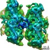
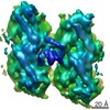
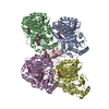
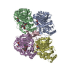
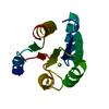
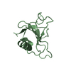
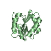
ジャーナル: Nat Struct Mol Biol / 年: 2017 タイトル: A structural model for microtubule minus-end recognition and protection by CAMSAP proteins. 著者: Joseph Atherton / Kai Jiang / Marcel M Stangier / Yanzhang Luo / Shasha Hua / Klaartje Houben / Jolien J E van Hooff / Agnel-Praveen Joseph / Guido Scarabelli / Barry J Grant / Anthony J ...著者: Joseph Atherton / Kai Jiang / Marcel M Stangier / Yanzhang Luo / Shasha Hua / Klaartje Houben / Jolien J E van Hooff / Agnel-Praveen Joseph / Guido Scarabelli / Barry J Grant / Anthony J Roberts / Maya Topf / Michel O Steinmetz / Marc Baldus / Carolyn A Moores / Anna Akhmanova / 要旨: CAMSAP and Patronin family members regulate microtubule minus-end stability and localization and thus organize noncentrosomal microtubule networks, which are essential for cell division, polarization ...CAMSAP and Patronin family members regulate microtubule minus-end stability and localization and thus organize noncentrosomal microtubule networks, which are essential for cell division, polarization and differentiation. Here, we found that the CAMSAP C-terminal CKK domain is widely present among eukaryotes and autonomously recognizes microtubule minus ends. Through a combination of structural approaches, we uncovered how mammalian CKK binds between two tubulin dimers at the interprotofilament interface on the outer microtubule surface. In vitro reconstitution assays combined with high-resolution fluorescence microscopy and cryo-electron tomography suggested that CKK preferentially associates with the transition zone between curved protofilaments and the regular microtubule lattice. We propose that minus-end-specific features of the interprotofilament interface at this site serve as the basis for CKK's minus-end preference. The steric clash between microtubule-bound CKK and kinesin motors explains how CKK protects microtubule minus ends against kinesin-13-induced depolymerization and thus controls the stability of free microtubule minus ends.
解像度: 1.4→68.14 Å / Cor.coef. Fo:Fc: 0.975 / Cor.coef. Fo:Fc free: 0.967 / SU B: 3.207 / SU ML: 0.051 / 交差検証法: THROUGHOUT / σ(F): 0 / ESU R: 0.051 / ESU R Free: 0.051 / 立体化学のターゲット値: MAXIMUM LIKELIHOOD 詳細: HYDROGENS HAVE BEEN ADDED IN THE RIDING POSITIONS U VALUES : WITH TLS ADDED
Rfactor
反射数
%反射
Selection details
Rfree
0.1916
1469
5 %
RANDOM
Rwork
0.162
-
-
-
obs
0.1636
28088
100 %
-
溶媒の処理
イオンプローブ半径: 0.8 Å / 減衰半径: 0.8 Å / VDWプローブ半径: 1.2 Å / 溶媒モデル: MASK
 ムービー
ムービー コントローラー


 データを開く
 基本情報
基本情報 要素
要素 キーワード
キーワード 機能・相同性情報
 X線回折 /
  データ登録者
 スイス, 2件
  引用
引用


 構造の表示
構造の表示 ダウンロードとリンク
 その他のダウンロード
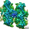


 PDBj
PDBj

 集合体
集合体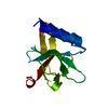

 分子量: 18.015 Da / 分子数: 67 / 由来タイプ: 天然 / 式: H2O
 試料調製
試料調製 解析
解析