 Movie
Movie Controller

[English] 日本語
 Yorodumi
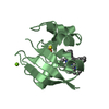
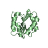
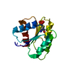
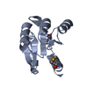
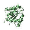
- PDB-6m8o: Crystal structure of the receiver domain of LytR from Staphylococ... -
+ Open data

- Basic information
| Entry | Database: PDB / ID: 6m8o | ||||||
|---|---|---|---|---|---|---|---|
| Title | Crystal structure of the receiver domain of LytR from Staphylococcus aureus | ||||||
 Components Components | DNA-binding response regulator | ||||||
 Keywords Keywords | SIGNALING PROTEIN / Two-Component System / Response Regulator / Receiver Domain | ||||||
| Function / homology |  Function and homology information Function and homology informationphosphorelay response regulator activity / protein-DNA complex / transcription cis-regulatory region binding / regulation of DNA-templated transcription / DNA binding / cytosol Similarity search - Function | ||||||
| Biological species |   Staphylococcus aureus (bacteria) Staphylococcus aureus (bacteria) | ||||||
| Method |  X-RAY DIFFRACTION / SYNCHROTRON / MOLECULAR REPLACEMENT / Resolution: 2.5 Å X-RAY DIFFRACTION / SYNCHROTRON / MOLECULAR REPLACEMENT / Resolution: 2.5 Å | ||||||
 Authors Authors | Shala-Lawrence, A. / Audette, G.F. | ||||||
| Funding support |  Canada, 1items Canada, 1items
| ||||||
 Citation Citation | Journal: To Be Published Title: Crystal structure of the receiver domain of LytR from Staphylococcus aureus Authors: Shala-Lawrence, A. / Audette, G.F. #1: Journal: Acta Crystallogr. Sect. F Struct. Biol. Cryst. Commun. Year: 2013 Title: Expression, purification, crystallization and preliminary X-ray analysis of the receiver domain of Staphylococcus aureus LytR protein. Authors: Shala, A. / Patel, K.H. / Golemi-Kotra, D. / Audette, G.F. | ||||||
| History |
|
- Structure visualization
| Structure viewer | Molecule: MolmilJmol/JSmol |
|---|
- Downloads & links
-Download
| PDBx/mmCIF format | 6m8o.cif.gz | 63.5 KB | Display | PDBx/mmCIF format |
|---|---|---|---|---|
| PDB format | pdb6m8o.ent.gz | 46.8 KB | Display | PDB format |
| PDBx/mmJSON format | 6m8o.json.gz | Tree view | PDBx/mmJSON format | |
| Others |  Other downloads Other downloads |
-Validation report
| Arichive directory | https://data.pdbj.org/pub/pdb/validation_reports/m8/6m8oftp://data.pdbj.org/pub/pdb/validation_reports/m8/6m8o | HTTPS FTP |
|---|
-Related structure data
| Related structure data |  2qvoS S: Starting model for refinement |
|---|---|
| Similar structure data |


-Links
 PDBj
PDBj

- Assembly
| Deposited unit | 
| ||||||||
|---|---|---|---|---|---|---|---|---|---|
| 1 |
| ||||||||
| 2 | 
| ||||||||
| Unit cell |
|
-Components
| #1: Protein | Mass: 15041.998 Da / Num. of mol.: 1 / Fragment: Response regulatory domain, residues 1-134 Source method: isolated from a genetically manipulated source Source: (gene. exp.) Staphylococcus aureus (bacteria) / Gene: C3B39_09020, EP54_06415, EQ90_12235, HMPREF3211_02791 / Production host: |
|---|---|
| #2: Chemical | ChemComp-SO4 /   Mass: 96.063 Da / Num. of mol.: 1 / Source method: obtained synthetically / Formula: SO4 Mass: 96.063 Da / Num. of mol.: 1 / Source method: obtained synthetically / Formula: SO4 |
| #3: Water | ChemComp-HOH /  Mass: 18.015 Da / Num. of mol.: 13 / Source method: isolated from a natural source / Formula: H2O Mass: 18.015 Da / Num. of mol.: 13 / Source method: isolated from a natural source / Formula: H2O |
-Experimental details
-Experiment
| Experiment | Method: X-RAY DIFFRACTION / Number of used crystals: 1 |
|---|
- Sample preparation
| Crystal | Density Matthews: 5.44 Å3/Da / Density % sol: 77.39 % |
|---|---|
| Crystal grow | Temperature: 277 K / Method: vapor diffusion, hanging drop / pH: 6.5 Details: 100 mM MES (pH 6.5), 1.6 M magnesium sulfate heptahydrate |
-Data collection
| Diffraction | Mean temperature: 100 K |
|---|---|
| Diffraction source | Source: SYNCHROTRON / Site: CLSI / Beamline: 08ID-1 / Wavelength: 0.9795 Å |
| Detector | Type: RAYONIX MX-300 / Detector: CCD / Date: Aug 15, 2013 |
| Radiation | Protocol: SINGLE WAVELENGTH / Monochromatic (M) / Laue (L): M / Scattering type: x-ray |
| Radiation wavelength | Wavelength: 0.9795 Å / Relative weight: 1 |
| Reflection | Resolution: 2.5→42.66 Å / Num. obs: 12211 / % possible obs: 99.9 % / Redundancy: 20.9 % / Biso Wilson estimate: 80.2 Å2 / CC1/2: 0.998 / Rmerge(I) obs: 0.087 / Rpim(I) all: 0.026 / Rrim(I) all: 0.089 / Χ2: 0.98 / Net I/σ(I): 20.3 |
| Reflection shell | Resolution: 2.5→2.6 Å / Redundancy: 21.5 % / Rmerge(I) obs: 2.607 / Mean I/σ(I) obs: 1.8 / Num. unique obs: 1323 / CC1/2: 0.91 / Rpim(I) all: 0.781 / Rrim(I) all: 2.669 / Χ2: 0.93 / % possible all: 99.7 |
- Processing
| Software |
| ||||||||||||||||||||||||||||||||||||||||||||||||||||||||||||||||||||||||||||||||||||||||||||||||||||||||||||||||||||||||||||||||||||||||||||||||||||||||||||||||||||||||||||||||||||||
|---|---|---|---|---|---|---|---|---|---|---|---|---|---|---|---|---|---|---|---|---|---|---|---|---|---|---|---|---|---|---|---|---|---|---|---|---|---|---|---|---|---|---|---|---|---|---|---|---|---|---|---|---|---|---|---|---|---|---|---|---|---|---|---|---|---|---|---|---|---|---|---|---|---|---|---|---|---|---|---|---|---|---|---|---|---|---|---|---|---|---|---|---|---|---|---|---|---|---|---|---|---|---|---|---|---|---|---|---|---|---|---|---|---|---|---|---|---|---|---|---|---|---|---|---|---|---|---|---|---|---|---|---|---|---|---|---|---|---|---|---|---|---|---|---|---|---|---|---|---|---|---|---|---|---|---|---|---|---|---|---|---|---|---|---|---|---|---|---|---|---|---|---|---|---|---|---|---|---|---|---|---|---|---|
| Refinement | Method to determine structure: MOLECULAR REPLACEMENT Starting model: 2qvo Resolution: 2.5→42.7 Å / Cor.coef. Fo:Fc: 0.959 / Cor.coef. Fo:Fc free: 0.944 / SU B: 18.513 / SU ML: 0.161 / Cross valid method: THROUGHOUT / ESU R: 0.178 / ESU R Free: 0.173 / Stereochemistry target values: MAXIMUM LIKELIHOOD / Details: HYDROGENS HAVE BEEN ADDED IN THE RIDING POSITIONS
| ||||||||||||||||||||||||||||||||||||||||||||||||||||||||||||||||||||||||||||||||||||||||||||||||||||||||||||||||||||||||||||||||||||||||||||||||||||||||||||||||||||||||||||||||||||||
| Solvent computation | Ion probe radii: 0.8 Å / Shrinkage radii: 0.8 Å / VDW probe radii: 1.2 Å / Solvent model: MASK | ||||||||||||||||||||||||||||||||||||||||||||||||||||||||||||||||||||||||||||||||||||||||||||||||||||||||||||||||||||||||||||||||||||||||||||||||||||||||||||||||||||||||||||||||||||||
| Displacement parameters | Biso mean: 91.566 Å2
| ||||||||||||||||||||||||||||||||||||||||||||||||||||||||||||||||||||||||||||||||||||||||||||||||||||||||||||||||||||||||||||||||||||||||||||||||||||||||||||||||||||||||||||||||||||||
| Refinement step | Cycle: 1 / Resolution: 2.5→42.7 Å
| ||||||||||||||||||||||||||||||||||||||||||||||||||||||||||||||||||||||||||||||||||||||||||||||||||||||||||||||||||||||||||||||||||||||||||||||||||||||||||||||||||||||||||||||||||||||
| Refine LS restraints |
|