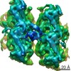
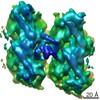
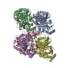
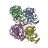
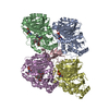
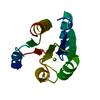
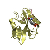
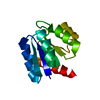
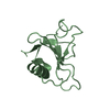
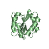
Journal: Nat Struct Mol Biol / Year: 2017 Title: A structural model for microtubule minus-end recognition and protection by CAMSAP proteins. Authors: Joseph Atherton / Kai Jiang / Marcel M Stangier / Yanzhang Luo / Shasha Hua / Klaartje Houben / Jolien J E van Hooff / Agnel-Praveen Joseph / Guido Scarabelli / Barry J Grant / Anthony J ...Authors: Joseph Atherton / Kai Jiang / Marcel M Stangier / Yanzhang Luo / Shasha Hua / Klaartje Houben / Jolien J E van Hooff / Agnel-Praveen Joseph / Guido Scarabelli / Barry J Grant / Anthony J Roberts / Maya Topf / Michel O Steinmetz / Marc Baldus / Carolyn A Moores / Anna Akhmanova / Abstract: CAMSAP and Patronin family members regulate microtubule minus-end stability and localization and thus organize noncentrosomal microtubule networks, which are essential for cell division, polarization ...CAMSAP and Patronin family members regulate microtubule minus-end stability and localization and thus organize noncentrosomal microtubule networks, which are essential for cell division, polarization and differentiation. Here, we found that the CAMSAP C-terminal CKK domain is widely present among eukaryotes and autonomously recognizes microtubule minus ends. Through a combination of structural approaches, we uncovered how mammalian CKK binds between two tubulin dimers at the interprotofilament interface on the outer microtubule surface. In vitro reconstitution assays combined with high-resolution fluorescence microscopy and cryo-electron tomography suggested that CKK preferentially associates with the transition zone between curved protofilaments and the regular microtubule lattice. We propose that minus-end-specific features of the interprotofilament interface at this site serve as the basis for CKK's minus-end preference. The steric clash between microtubule-bound CKK and kinesin motors explains how CKK protects microtubule minus ends against kinesin-13-induced depolymerization and thus controls the stability of free microtubule minus ends.
Resolution: 1.4→68.14 Å / Cor.coef. Fo:Fc: 0.975 / Cor.coef. Fo:Fc free: 0.967 / SU B: 3.207 / SU ML: 0.051 / Cross valid method: THROUGHOUT / σ(F): 0 / ESU R: 0.051 / ESU R Free: 0.051 / Stereochemistry target values: MAXIMUM LIKELIHOOD Details: HYDROGENS HAVE BEEN ADDED IN THE RIDING POSITIONS U VALUES : WITH TLS ADDED
Rfactor
Num. reflection
% reflection
Selection details
Rfree
0.1916
1469
5 %
RANDOM
Rwork
0.162
-
-
-
obs
0.1636
28088
100 %
-
Solvent computation
Ion probe radii: 0.8 Å / Shrinkage radii: 0.8 Å / VDW probe radii: 1.2 Å / Solvent model: MASK
In the structure databanks used in Yorodumi, some data are registered as the other names, "COVID-19 virus" and "2019-nCoV". Here are the details of the virus and the list of structure data.
Jan 31, 2019. EMDB accession codes are about to change! (news from PDBe EMDB page)
EMDB accession codes are about to change! (news from PDBe EMDB page)
The allocation of 4 digits for EMDB accession codes will soon come to an end. Whilst these codes will remain in use, new EMDB accession codes will include an additional digit and will expand incrementally as the available range of codes is exhausted. The current 4-digit format prefixed with “EMD-” (i.e. EMD-XXXX) will advance to a 5-digit format (i.e. EMD-XXXXX), and so on. It is currently estimated that the 4-digit codes will be depleted around Spring 2019, at which point the 5-digit format will come into force.
The EM Navigator/Yorodumi systems omit the EMD- prefix.
Related info.:Q: What is EMD? / ID/Accession-code notation in Yorodumi/EM Navigator
Yorodumi is a browser for structure data from EMDB, PDB, SASBDB, etc.
This page is also the successor to EM Navigator detail page, and also detail information page/front-end page for Omokage search.
The word "yorodu" (or yorozu) is an old Japanese word meaning "ten thousand". "mi" (miru) is to see.
Related info.:EMDB / PDB / SASBDB / Comparison of 3 databanks / Yorodumi Search / Aug 31, 2016. New EM Navigator & Yorodumi / Yorodumi Papers / Jmol/JSmol / Function and homology information / Changes in new EM Navigator and Yorodumi
 Movie
Movie Controller


 Open data
 Basic information
 Components
 Keywords
 Function and homology information
 X-RAY DIFFRACTION /
  Authors
 Switzerland, 2items
  Citation


 Structure visualization
 Downloads & links
 Other downloads
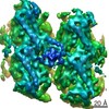


 PDBj
PDBj

 Assembly
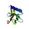

 Mass: 18.015 Da / Num. of mol.: 67 / Source method: isolated from a natural source / Formula: H2O
 Sample preparation
 Processing