 Movie
Movie Controller

[English] 日本語
 Yorodumi
- PDB-5l8r: The structure of plant photosystem I super-complex at 2.6 angstro... -
+ Open data

- Basic information
| Entry | Database: PDB / ID: 5l8r | ||||||||||||
|---|---|---|---|---|---|---|---|---|---|---|---|---|---|
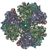
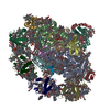
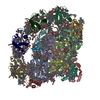
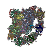
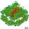
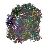
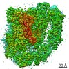
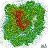
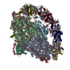
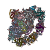
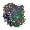
| Title | The structure of plant photosystem I super-complex at 2.6 angstrom resolution. | ||||||||||||
 Components Components |
| ||||||||||||
 Keywords Keywords |  OXIDOREDUCTASE / photosynthesis / membrane complex / chlorophyll / light harvesting OXIDOREDUCTASE / photosynthesis / membrane complex / chlorophyll / light harvesting | ||||||||||||
| Function / homology |  Function and homology information Function and homology informationphotosynthetic NADP+ reduction / photosystem I stabilization / chloroplast photosystem I / chloroplast membrane / chloroplast thylakoid / photosynthesis, light harvesting / thylakoid / chloroplast thylakoid lumen / photosynthesis, light harvesting in photosystem I / photosystem I reaction center ...photosynthetic NADP+ reduction / photosystem I stabilization / chloroplast photosystem I / chloroplast membrane / chloroplast thylakoid / photosynthesis, light harvesting / thylakoid / chloroplast thylakoid lumen / photosynthesis, light harvesting in photosystem I / photosystem I reaction center / photosystem I / chloroplast envelope / photosystem I / photosystem II / photosynthetic electron transport in photosystem I / chlorophyll binding / chloroplast thylakoid membrane / response to light stimulus / photosynthesis / chloroplast / 4 iron, 4 sulfur cluster binding / electron transfer activity / oxidoreductase activity / protein stabilization / magnesium ion binding / metal ion binding / nucleusSimilarity search - Function | ||||||||||||
| Biological species |   Pisum sativum (garden pea) Pisum sativum (garden pea) | ||||||||||||
| Method | X-RAY DIFFRACTION / SYNCHROTRON / SAD / Resolution: 2.6 Å | ||||||||||||
 Authors Authors | Mazor, Y. / Borovikova, A. / Caspy, I. / Nelson, N. | ||||||||||||
| Funding support |  Israel, 3items Israel, 3items
| ||||||||||||
 Citation Citation | Journal: Nat Plants / Year: 2017 Title: Structure of the plant photosystem I supercomplex at 2.6 angstrom resolution. Authors: Mazor, Y. / Borovikova, A. / Caspy, I. / Nelson, N. | ||||||||||||
| History |
|
- Structure visualization
| Structure viewer | Molecule: MolmilJmol/JSmol |
|---|
- Downloads & links
-Download
| PDBx/mmCIF format | 5l8r.cif.gz | 1.9 MB | Display | PDBx/mmCIF format |
|---|---|---|---|---|
| PDB format | pdb5l8r.ent.gz | 1.7 MB | Display | PDB format |
| PDBx/mmJSON format | 5l8r.json.gz | Tree view | PDBx/mmJSON format | |
| Others |  Other downloads Other downloads |
-Validation report
| Arichive directory | https://data.pdbj.org/pub/pdb/validation_reports/l8/5l8rftp://data.pdbj.org/pub/pdb/validation_reports/l8/5l8r | HTTPS FTP |
|---|
-Related structure data
| Related structure data | |
|---|---|
| Similar structure data |


-Links
 PDBj
PDBj


- Assembly
| Deposited unit | 
| ||||||||
|---|---|---|---|---|---|---|---|---|---|
| 1 |
| ||||||||
| Unit cell |
|
-Components
-Protein , 5 types, 5 molecules 12CDG
| #1: Protein | Mass: 21335.439 Da / Num. of mol.: 1 / Source method: isolated from a natural source / Source: (natural) Pisum sativum (garden pea) / References: UniProt: E1C9L2*PLUS |
|---|---|
| #2: Protein | Mass: 28912.719 Da / Num. of mol.: 1 / Source method: isolated from a natural source / Source: (natural) Pisum sativum (garden pea) / References: UniProt: Q41038 |
| #7: Protein | / 9 kDa polypeptide / PSI-C / Photosystem I subunit VII / PsaC Mass: 8991.474 Da / Num. of mol.: 1 / Source method: isolated from a natural source / Source: (natural) Pisum sativum (garden pea) / References: UniProt: P10793, photosystem I |
| #8: Protein | Mass: 16041.408 Da / Num. of mol.: 1 / Source method: isolated from a natural source / Source: (natural) Pisum sativum (garden pea) / References: UniProt: E1C9K8*PLUS |
| #11: Protein | Mass: 10678.027 Da / Num. of mol.: 1 / Source method: isolated from a natural source / Source: (natural) Pisum sativum (garden pea) / References: UniProt: Q9S7N7*PLUS |
-Chlorophyll a-b binding protein ... , 2 types, 2 molecules 34
| #3: Protein | Mass: 29634.801 Da / Num. of mol.: 1 / Source method: isolated from a natural source / Source: (natural) Pisum sativum (garden pea) / References: UniProt: Q32904 |
|---|---|
| #4: Protein | Mass: 21994.029 Da / Num. of mol.: 1 / Source method: isolated from a natural source / Source: (natural) Pisum sativum (garden pea) / References: UniProt: Q9SQL2 |
-Photosystem I P700 chlorophyll a apoprotein ... , 2 types, 2 molecules AB
| #5: Protein | / PSI-A / PsaA Mass: 84265.539 Da / Num. of mol.: 1 / Source method: isolated from a natural source / Source: (natural) Pisum sativum (garden pea) / References: UniProt: P05310, photosystem I |
|---|---|
| #6: Protein | / PSI-B / PsaB Mass: 82512.938 Da / Num. of mol.: 1 / Source method: isolated from a natural source / Source: (natural) Pisum sativum (garden pea) / References: UniProt: A0A0F6NGI2, photosystem I |
-Putative uncharacterized ... , 2 types, 2 molecules EL
| #9: Protein | Mass: 7479.422 Da / Num. of mol.: 1 / Source method: isolated from a natural source / Source: (natural) Pisum sativum (garden pea) / References: UniProt: E1C9K6 |
|---|---|
| #16: Protein | Mass: 16564.977 Da / Num. of mol.: 1 / Source method: isolated from a natural source / Source: (natural) Pisum sativum (garden pea) / References: UniProt: E1C9L1 |
-Photosystem I reaction center subunit ... , 5 types, 5 molecules FHIJK
| #10: Protein | Mass: 17238.000 Da / Num. of mol.: 1 / Source method: isolated from a natural source / Source: (natural) Pisum sativum (garden pea) / References: UniProt: A0A0M3KL12 |
|---|---|
| #12: Protein | Mass: 9491.742 Da / Num. of mol.: 1 / Source method: isolated from a natural source / Source: (natural) Pisum sativum (garden pea) / References: UniProt: A0A0M3KL10 |
| #13: Protein/peptide | / PSI-I Mass: 4474.527 Da / Num. of mol.: 1 / Source method: isolated from a natural source / Source: (natural) Pisum sativum (garden pea) / References: UniProt: P17227 |
| #14: Protein/peptide | / PSI-J Mass: 4767.609 Da / Num. of mol.: 1 / Source method: isolated from a natural source / Source: (natural) Pisum sativum (garden pea) / References: UniProt: D5MAL3 |
| #15: Protein | Mass: 7988.296 Da / Num. of mol.: 1 / Source method: isolated from a natural source / Source: (natural) Pisum sativum (garden pea) / References: UniProt: E1C9L3 |
-Sugars , 2 types, 15 molecules 

| #24: Sugar | ChemComp-LMT /  Type: D-saccharide / Mass: 510.615 Da / Num. of mol.: 10 / Source method: obtained synthetically / Formula: C24H46O11 / Comment: detergent*YM Type: D-saccharide / Mass: 510.615 Da / Num. of mol.: 10 / Source method: obtained synthetically / Formula: C24H46O11 / Comment: detergent*YM#26: Sugar | ChemComp-DGD /  Type: saccharideCarbohydrate / Mass: 949.299 Da / Num. of mol.: 5 / Source method: obtained synthetically / Formula: C51H96O15 Type: saccharideCarbohydrate / Mass: 949.299 Da / Num. of mol.: 5 / Source method: obtained synthetically / Formula: C51H96O15 |
|---|
-Non-polymers , 13 types, 436 molecules 


| #17: Chemical | ChemComp-LUT / ( Lutein Mass: 568.871 Da / Num. of mol.: 7 / Source method: obtained synthetically / Formula: C40H56O2 Mass: 568.871 Da / Num. of mol.: 7 / Source method: obtained synthetically / Formula: C40H56O2#18: Chemical | ChemComp-BCR / Β-Carotene Mass: 536.873 Da / Num. of mol.: 27 / Source method: obtained synthetically / Formula: C40H56 Mass: 536.873 Da / Num. of mol.: 27 / Source method: obtained synthetically / Formula: C40H56#19: Chemical | ChemComp-CLA / Chlorophyll a Mass: 893.489 Da / Num. of mol.: 142 / Source method: obtained synthetically / Formula: C55H72MgN4O5 Mass: 893.489 Da / Num. of mol.: 142 / Source method: obtained synthetically / Formula: C55H72MgN4O5#20: Chemical | ChemComp-CHL / Chlorophyll b Mass: 907.472 Da / Num. of mol.: 13 / Source method: obtained synthetically / Formula: C55H70MgN4O6 Mass: 907.472 Da / Num. of mol.: 13 / Source method: obtained synthetically / Formula: C55H70MgN4O6#21: Chemical | ChemComp-LHG / Phosphatidylglycerol Mass: 722.970 Da / Num. of mol.: 7 / Source method: obtained synthetically / Formula: C38H75O10P / Comment: phospholipid*YM Mass: 722.970 Da / Num. of mol.: 7 / Source method: obtained synthetically / Formula: C38H75O10P / Comment: phospholipid*YM#22: Chemical | ChemComp-LMG /  Mass: 787.158 Da / Num. of mol.: 20 / Source method: obtained synthetically / Formula: C45H86O10 Mass: 787.158 Da / Num. of mol.: 20 / Source method: obtained synthetically / Formula: C45H86O10#23: Chemical | Violaxanthin Mass: 600.870 Da / Num. of mol.: 2 / Source method: obtained synthetically / Formula: C40H56O4 Mass: 600.870 Da / Num. of mol.: 2 / Source method: obtained synthetically / Formula: C40H56O4#25: Chemical |  Mass: 40.078 Da / Num. of mol.: 2 / Source method: obtained synthetically / Formula: Ca Mass: 40.078 Da / Num. of mol.: 2 / Source method: obtained synthetically / Formula: Ca#27: Chemical | ChemComp-CL0 / | Chlorophyll a Mass: 893.489 Da / Num. of mol.: 1 / Source method: obtained synthetically / Formula: C55H72MgN4O5 Mass: 893.489 Da / Num. of mol.: 1 / Source method: obtained synthetically / Formula: C55H72MgN4O5#28: Chemical | Iron–sulfur cluster Mass: 351.640 Da / Num. of mol.: 3 / Source method: obtained synthetically / Formula: Fe4S4 Mass: 351.640 Da / Num. of mol.: 3 / Source method: obtained synthetically / Formula: Fe4S4#29: Chemical | Phytomenadione Mass: 450.696 Da / Num. of mol.: 2 / Source method: obtained synthetically / Formula: C31H46O2 Mass: 450.696 Da / Num. of mol.: 2 / Source method: obtained synthetically / Formula: C31H46O2#30: Chemical | ChemComp-ZEX / ( |  Mass: 568.871 Da / Num. of mol.: 1 / Source method: obtained synthetically / Formula: C40H56O2 Mass: 568.871 Da / Num. of mol.: 1 / Source method: obtained synthetically / Formula: C40H56O2#31: Water | ChemComp-HOH / | WaterMass: 18.015 Da / Num. of mol.: 209 / Source method: isolated from a natural source / Formula: H2O |
|---|
-Experimental details
-Experiment
| Experiment | Method: X-RAY DIFFRACTION / Number of used crystals: 1 |
|---|
- Sample preparation
| Crystal grow | Temperature: 277 K / Method: vapor diffusion, sitting drop / pH: 8.75 Details: 30mM Tris-Hcl pH8.75, 50mM KH2PO4, 0.03% Octyl Glucose Neopentyl Glycol, 14% PEG400, 2mM glutathione |
|---|
-Data collection
| Diffraction | Mean temperature: 100 K |
|---|---|
| Diffraction source | Source: SYNCHROTRON / Site: SLS  / Beamline: X06DA / Wavelength: 1 Å / Beamline: X06DA / Wavelength: 1 Å |
| Detector | Type: DECTRIS PILATUS3 6M / Detector: PIXEL / Date: Jun 1, 2015 |
| Radiation | Monochromator: Si(111) / Protocol: SINGLE WAVELENGTH / Monochromatic (M) / Laue (L): M / Scattering type: x-ray |
| Radiation wavelength | Wavelength: 1 Å / Relative weight: 1 |
| Reflection | Resolution: 2.6→50 Å / Num. obs: 248807 / % possible obs: 99.9 % / Redundancy: 84.6 % / CC1/2: 0.997 / Net I/σ(I): 9.8 |
| Reflection shell | Resolution: 2.6→2.64 Å / Redundancy: 83.9 % / Mean I/σ(I) obs: 1.5 / % possible all: 99.4 |
- Processing
| Software |
| |||||||||||||||||||||||||||||||||||||||||||||||||||||||||||||||||||||||||||||||||||||||||||||||||||||||||||||||||||||||||||||||||||||||||||||||||||||||||||||||||||||||||||||||||||||||||||||||||||||||||||||||||||||||||
|---|---|---|---|---|---|---|---|---|---|---|---|---|---|---|---|---|---|---|---|---|---|---|---|---|---|---|---|---|---|---|---|---|---|---|---|---|---|---|---|---|---|---|---|---|---|---|---|---|---|---|---|---|---|---|---|---|---|---|---|---|---|---|---|---|---|---|---|---|---|---|---|---|---|---|---|---|---|---|---|---|---|---|---|---|---|---|---|---|---|---|---|---|---|---|---|---|---|---|---|---|---|---|---|---|---|---|---|---|---|---|---|---|---|---|---|---|---|---|---|---|---|---|---|---|---|---|---|---|---|---|---|---|---|---|---|---|---|---|---|---|---|---|---|---|---|---|---|---|---|---|---|---|---|---|---|---|---|---|---|---|---|---|---|---|---|---|---|---|---|---|---|---|---|---|---|---|---|---|---|---|---|---|---|---|---|---|---|---|---|---|---|---|---|---|---|---|---|---|---|---|---|---|---|---|---|---|---|---|---|---|---|---|---|---|---|---|---|---|
| Refinement | Method to determine structure: SAD / Resolution: 2.6→39.906 Å / SU ML: 0.4 / Cross valid method: FREE R-VALUE / σ(F): 1.33 / Phase error: 28.27 / Stereochemistry target values: MLHL
| |||||||||||||||||||||||||||||||||||||||||||||||||||||||||||||||||||||||||||||||||||||||||||||||||||||||||||||||||||||||||||||||||||||||||||||||||||||||||||||||||||||||||||||||||||||||||||||||||||||||||||||||||||||||||
| Solvent computation | Shrinkage radii: 0.9 Å / VDW probe radii: 1.11 Å / Solvent model: FLAT BULK SOLVENT MODEL | |||||||||||||||||||||||||||||||||||||||||||||||||||||||||||||||||||||||||||||||||||||||||||||||||||||||||||||||||||||||||||||||||||||||||||||||||||||||||||||||||||||||||||||||||||||||||||||||||||||||||||||||||||||||||
| Refinement step | Cycle: LAST / Resolution: 2.6→39.906 Å
| |||||||||||||||||||||||||||||||||||||||||||||||||||||||||||||||||||||||||||||||||||||||||||||||||||||||||||||||||||||||||||||||||||||||||||||||||||||||||||||||||||||||||||||||||||||||||||||||||||||||||||||||||||||||||
| Refine LS restraints |
| |||||||||||||||||||||||||||||||||||||||||||||||||||||||||||||||||||||||||||||||||||||||||||||||||||||||||||||||||||||||||||||||||||||||||||||||||||||||||||||||||||||||||||||||||||||||||||||||||||||||||||||||||||||||||
| LS refinement shell |
| |||||||||||||||||||||||||||||||||||||||||||||||||||||||||||||||||||||||||||||||||||||||||||||||||||||||||||||||||||||||||||||||||||||||||||||||||||||||||||||||||||||||||||||||||||||||||||||||||||||||||||||||||||||||||
| Refinement TLS params. | Method: refined / Origin x: 15.038 Å / Origin y: 7.6729 Å / Origin z: 64.5628 Å
| |||||||||||||||||||||||||||||||||||||||||||||||||||||||||||||||||||||||||||||||||||||||||||||||||||||||||||||||||||||||||||||||||||||||||||||||||||||||||||||||||||||||||||||||||||||||||||||||||||||||||||||||||||||||||
| Refinement TLS group | Selection details: all |
