 Movie
Movie Controller


+ Open data

- Basic information
| Entry | Database: PDB / ID: 6l35 | ||||||||||||||||||||||||||||||
|---|---|---|---|---|---|---|---|---|---|---|---|---|---|---|---|---|---|---|---|---|---|---|---|---|---|---|---|---|---|---|---|
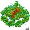
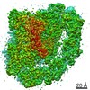
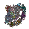
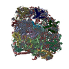
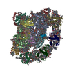
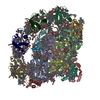
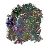
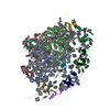
| Title | PSI-LHCI Supercomplex from Physcometrella patens | ||||||||||||||||||||||||||||||
 Components Components |
| ||||||||||||||||||||||||||||||
 Keywords Keywords | STRUCTURAL PROTEIN / Cryo-EM / PSI-LHCI / Physcomitrella patens | ||||||||||||||||||||||||||||||
| Function / homology |  Function and homology information Function and homology informationphotosystem I antenna complex / pigment binding / response to low light intensity stimulus / response to high light intensity / photosynthesis, light harvesting in photosystem I / photosynthesis, light harvesting / chloroplast thylakoid lumen / photosystem I reaction center / photosystem I / photosynthetic electron transport in photosystem I ...photosystem I antenna complex / pigment binding / response to low light intensity stimulus / response to high light intensity / photosynthesis, light harvesting in photosystem I / photosynthesis, light harvesting / chloroplast thylakoid lumen / photosystem I reaction center / photosystem I / photosynthetic electron transport in photosystem I / photosystem I / photosystem II / chlorophyll binding / chloroplast thylakoid membrane / membrane => GO:0016020 / response to light stimulus / photosynthesis / response to cold / chloroplast / 4 iron, 4 sulfur cluster binding / electron transfer activity / oxidoreductase activity / serine-type endopeptidase activity / protein domain specific binding / magnesium ion binding / protein homodimerization activity / metal ion binding Similarity search - Function | ||||||||||||||||||||||||||||||
| Biological species |  Physcomitrium patens (plant) Physcomitrium patens (plant) | ||||||||||||||||||||||||||||||
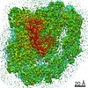
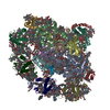
| Method | ELECTRON MICROSCOPY / single particle reconstruction / Resolution: 3.23 Å | ||||||||||||||||||||||||||||||
 Authors Authors | Zhao, L. / Yan, Q.J. / Qin, X.C. | ||||||||||||||||||||||||||||||
 Citation Citation | Journal: Cell Discov / Year: 2021 Title: Antenna arrangement and energy-transfer pathways of PSI-LHCI from the moss Physcomitrella patens. Authors: Qiujing Yan / Liang Zhao / Wenda Wang / Xiong Pi / Guangye Han / Jie Wang / Lingpeng Cheng / Yi-Kun He / Tingyun Kuang / Xiaochun Qin / Sen-Fang Sui / Jian-Ren Shen /   Abstract: Plants harvest light energy utilized for photosynthesis by light-harvesting complex I and II (LHCI and LHCII) surrounding photosystem I and II (PSI and PSII), respectively. During the evolution of ...Plants harvest light energy utilized for photosynthesis by light-harvesting complex I and II (LHCI and LHCII) surrounding photosystem I and II (PSI and PSII), respectively. During the evolution of green plants, moss is at an evolutionarily intermediate position from aquatic photosynthetic organisms to land plants, being the first photosynthetic organisms that landed. Here, we report the structure of the PSI-LHCI supercomplex from the moss Physcomitrella patens (Pp) at 3.23 Å resolution solved by cryo-electron microscopy. Our structure revealed that four Lhca subunits are associated with the PSI core in an order of Lhca1-Lhca5-Lhca2-Lhca3. This number is much decreased from 8 to 10, the number of subunits in most green algal PSI-LHCI, but the same as those of land plants. Although Pp PSI-LHCI has a similar structure as PSI-LHCI of land plants, it has Lhca5, instead of Lhca4, in the second position of Lhca, and several differences were found in the arrangement of chlorophylls among green algal, moss, and land plant PSI-LHCI. One chlorophyll, PsaF-Chl 305, which is found in the moss PSI-LHCI, is located at the gap region between the two middle Lhca subunits and the PSI core, and therefore may make the excitation energy transfer from LHCI to the core more efficient than that of land plants. On the other hand, energy-transfer paths at the two side Lhca subunits are relatively conserved. These results provide a structural basis for unravelling the mechanisms of light-energy harvesting and transfer in the moss PSI-LHCI, as well as important clues on the changes of PSI-LHCI after landing. | ||||||||||||||||||||||||||||||
| History |
|
- Structure visualization
| Movie |
Movie viewer |
|---|---|
| Structure viewer | Molecule: MolmilJmol/JSmol |
- Downloads & links
-Download
| PDBx/mmCIF format | 6l35.cif.gz | 787.1 KB | Display | PDBx/mmCIF format |
|---|---|---|---|---|
| PDB format | pdb6l35.ent.gz | 677.1 KB | Display | PDB format |
| PDBx/mmJSON format | 6l35.json.gz | Tree view | PDBx/mmJSON format | |
| Others |  Other downloads Other downloads |
-Validation report
| Arichive directory | https://data.pdbj.org/pub/pdb/validation_reports/l3/6l35ftp://data.pdbj.org/pub/pdb/validation_reports/l3/6l35 | HTTPS FTP |
|---|
-Related structure data
| Related structure data | 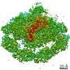 0821MC M: map data used to model this data C: citing same article ( |
|---|---|
| Similar structure data |
-Links
 PDBj
PDBj


- Assembly
| Deposited unit | 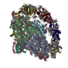
|
|---|---|
| 1 |
|
-Components
-Photosystem I P700 chlorophyll a apoprotein ... , 2 types, 2 molecules AB
| #1: Protein | Mass: 82313.422 Da / Num. of mol.: 1 / Source method: isolated from a natural source / Source: (natural) Physcomitrium patens (plant) / References: UniProt: Q8MFA3, photosystem I |
|---|---|
| #2: Protein | Mass: 82316.547 Da / Num. of mol.: 1 / Source method: isolated from a natural source / Source: (natural) Physcomitrium patens (plant) / References: UniProt: Q8MFA2, photosystem I |
-Protein , 6 types, 6 molecules CEFHKL
| #3: Protein | Mass: 8649.957 Da / Num. of mol.: 1 / Source method: isolated from a natural source / Source: (natural) Physcomitrium patens (plant) / References: UniProt: Q6YXQ2, photosystem I |
|---|---|
| #5: Protein | Mass: 6895.725 Da / Num. of mol.: 1 / Source method: isolated from a natural source / Source: (natural) Physcomitrium patens (plant) / References: UniProt: A0A2K1IKE2 |
| #6: Protein | Mass: 17465.363 Da / Num. of mol.: 1 / Source method: isolated from a natural source / Source: (natural) Physcomitrium patens (plant) / References: UniProt: A0A2K1J0L9 |
| #8: Protein | Mass: 9861.088 Da / Num. of mol.: 1 / Source method: isolated from a natural source / Source: (natural) Physcomitrium patens (plant) / References: UniProt: A9SL09 |
| #11: Protein | Mass: 7916.146 Da / Num. of mol.: 1 / Source method: isolated from a natural source / Source: (natural) Physcomitrium patens (plant) / References: UniProt: A9SFV4, UniProt: A0A2K1KU02*PLUS |
| #12: Protein | Mass: 16892.416 Da / Num. of mol.: 1 / Source method: isolated from a natural source / Source: (natural) Physcomitrium patens (plant) / References: UniProt: A9S7M7, UniProt: A0A2K1IAD0*PLUS |
-Predicted protein ... , 2 types, 2 molecules DG
| #4: Protein | Mass: 15654.927 Da / Num. of mol.: 1 / Source method: isolated from a natural source / Source: (natural) Physcomitrium patens (plant) / References: UniProt: A9SRC8, UniProt: A0A2K1JKF2*PLUS |
|---|---|
| #7: Protein | Mass: 10564.831 Da / Num. of mol.: 1 / Source method: isolated from a natural source / Source: (natural) Physcomitrium patens (plant) / References: UniProt: A9SJ10, UniProt: A0A2K1JC42*PLUS |
-Photosystem I reaction center subunit ... , 3 types, 3 molecules IJM
| #9: Protein/peptide | Mass: 3791.472 Da / Num. of mol.: 1 / Source method: isolated from a natural source / Source: (natural) Physcomitrium patens (plant) / References: UniProt: Q6YXR3 |
|---|---|
| #10: Protein/peptide | Mass: 4597.460 Da / Num. of mol.: 1 / Source method: isolated from a natural source / Source: (natural) Physcomitrium patens (plant) / References: UniProt: Q6YXM2 |
| #13: Protein/peptide | Mass: 3051.623 Da / Num. of mol.: 1 / Source method: isolated from a natural source / Source: (natural) Physcomitrium patens (plant) / References: UniProt: Q6YXK4 |
-Chlorophyll a-b binding protein, ... , 4 types, 4 molecules 2635
| #14: Protein | Mass: 22922.107 Da / Num. of mol.: 1 / Source method: isolated from a natural source / Source: (natural) Physcomitrium patens (plant) / References: UniProt: A0A2K1K0C7 |
|---|---|
| #15: Protein | Mass: 20761.584 Da / Num. of mol.: 1 / Source method: isolated from a natural source / Source: (natural) Physcomitrium patens (plant) / References: UniProt: A9T399, UniProt: A0A2K1JLZ3*PLUS |
| #16: Protein | Mass: 23250.502 Da / Num. of mol.: 1 / Source method: isolated from a natural source / Source: (natural) Physcomitrium patens (plant) / References: UniProt: A9TEM8, UniProt: A0A2K1IB10*PLUS |
| #17: Protein | Mass: 22445.592 Da / Num. of mol.: 1 / Source method: isolated from a natural source / Source: (natural) Physcomitrium patens (plant) / References: UniProt: A0A2K1KN29 |
-Sugars , 1 types, 1 molecules 
| #23: Sugar | ChemComp-DGD /  Type: saccharide / Mass: 949.299 Da / Num. of mol.: 1 / Source method: obtained synthetically / Formula: C51H96O15 Type: saccharide / Mass: 949.299 Da / Num. of mol.: 1 / Source method: obtained synthetically / Formula: C51H96O15 |
|---|
-Non-polymers , 9 types, 204 molecules 


| #18: Chemical | ChemComp-CLA /  Mass: 893.489 Da / Num. of mol.: 144 / Source method: obtained synthetically / Formula: C55H72MgN4O5 Mass: 893.489 Da / Num. of mol.: 144 / Source method: obtained synthetically / Formula: C55H72MgN4O5#19: Chemical |  Mass: 450.696 Da / Num. of mol.: 2 / Source method: obtained synthetically / Formula: C31H46O2 Mass: 450.696 Da / Num. of mol.: 2 / Source method: obtained synthetically / Formula: C31H46O2#20: Chemical | ChemComp-LHG /  Mass: 722.970 Da / Num. of mol.: 6 / Source method: obtained synthetically / Formula: C38H75O10P / Comment: phospholipid*YM Mass: 722.970 Da / Num. of mol.: 6 / Source method: obtained synthetically / Formula: C38H75O10P / Comment: phospholipid*YM#21: Chemical | ChemComp-BCR /  Mass: 536.873 Da / Num. of mol.: 26 / Source method: obtained synthetically / Formula: C40H56 Mass: 536.873 Da / Num. of mol.: 26 / Source method: obtained synthetically / Formula: C40H56#22: Chemical |  Mass: 351.640 Da / Num. of mol.: 3 / Source method: obtained synthetically / Formula: Fe4S4 Mass: 351.640 Da / Num. of mol.: 3 / Source method: obtained synthetically / Formula: Fe4S4#24: Chemical |  Mass: 787.158 Da / Num. of mol.: 3 / Source method: obtained synthetically / Formula: C45H86O10 Mass: 787.158 Da / Num. of mol.: 3 / Source method: obtained synthetically / Formula: C45H86O10#25: Chemical | ChemComp-CHL /  Mass: 907.472 Da / Num. of mol.: 12 / Source method: obtained synthetically / Formula: C55H70MgN4O6 Mass: 907.472 Da / Num. of mol.: 12 / Source method: obtained synthetically / Formula: C55H70MgN4O6#26: Chemical | ChemComp-LUT / (  Mass: 568.871 Da / Num. of mol.: 4 / Source method: obtained synthetically / Formula: C40H56O2 Mass: 568.871 Da / Num. of mol.: 4 / Source method: obtained synthetically / Formula: C40H56O2#27: Chemical | ChemComp-XAT / (  Mass: 600.870 Da / Num. of mol.: 4 / Source method: obtained synthetically / Formula: C40H56O4 Mass: 600.870 Da / Num. of mol.: 4 / Source method: obtained synthetically / Formula: C40H56O4 |
|---|
-Details
| Has ligand of interest | N |
|---|---|
| Has protein modification | Y |
-Experimental details
-Experiment
| Experiment | Method: ELECTRON MICROSCOPY |
|---|---|
| EM experiment | Aggregation state: PARTICLE / 3D reconstruction method: single particle reconstruction |
- Sample preparation
| Component | Name: PSI-LHCI / Type: COMPLEX / Source: NATURAL |
|---|---|
| Source (natural) | Organism: Physcomitrella patens (plant) |
| Buffer solution | pH: 7.4 |
| Specimen | Embedding applied: NO / Shadowing applied: NO / Staining applied: NO / Vitrification applied: NO |
- Electron microscopy imaging
| Experimental equipment |  Model: Titan Krios / Image courtesy: FEI Company |
|---|---|
| Microscopy | Model: FEI TITAN KRIOS |
| Electron gun | Electron source:  FIELD EMISSION GUN / Accelerating voltage: 300 kV / Illumination mode: FLOOD BEAM FIELD EMISSION GUN / Accelerating voltage: 300 kV / Illumination mode: FLOOD BEAM |
| Electron lens | Mode: BRIGHT FIELD |
| Image recording | Electron dose: 50 e/Å2 / Film or detector model: GATAN K2 SUMMIT (4k x 4k) |
- Processing
| Software | Name: PHENIX / Version: 1.17.1_3660: / Classification: refinement | ||||||||||||||||||||||||||||||||
|---|---|---|---|---|---|---|---|---|---|---|---|---|---|---|---|---|---|---|---|---|---|---|---|---|---|---|---|---|---|---|---|---|---|
| EM software |
| ||||||||||||||||||||||||||||||||
| CTF correction | Type: NONE | ||||||||||||||||||||||||||||||||
| Symmetry | Point symmetry: C1 (asymmetric) | ||||||||||||||||||||||||||||||||
| 3D reconstruction | Resolution: 3.23 Å / Resolution method: FSC 0.143 CUT-OFF / Num. of particles: 70288 / Symmetry type: POINT | ||||||||||||||||||||||||||||||||
| Refinement | Highest resolution: 3.23 Å | ||||||||||||||||||||||||||||||||
| Refine LS restraints |
|
