 Movie
Movie Controller

[English] 日本語
 Yorodumi
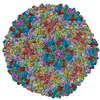
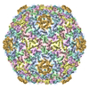
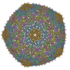
- PDB-5l35: Cryo-EM structure of bacteriophage Sf6 at 2.9 Angstrom resolution -
+ Open data

- Basic information
| Entry | Database: PDB / ID: 5l35 | ||||||
|---|---|---|---|---|---|---|---|
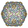
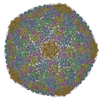
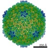
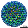
| Title | Cryo-EM structure of bacteriophage Sf6 at 2.9 Angstrom resolution | ||||||
 Components Components | Gene 5 protein | ||||||
 Keywords Keywords | VIRUS / phage / Sf6 | ||||||
| Function / homology | Major capsid protein Gp5 / P22 coat protein - gene protein 5 / Gene 5 protein Function and homology information Function and homology information | ||||||
| Biological species |  Shigella phage Sf6 (virus) Shigella phage Sf6 (virus) | ||||||
| Method | ELECTRON MICROSCOPY / single particle reconstruction / cryo EM / Resolution: 2.89 Å | ||||||
 Authors Authors | Zhao, H. / Tang, L. | ||||||
| Funding support |  United States, 1items United States, 1items
| ||||||
 Citation Citation | Journal: Proc Natl Acad Sci U S A / Year: 2017 Title: Structure of a headful DNA-packaging bacterial virus at 2.9 Å resolution by electron cryo-microscopy. Authors: Haiyan Zhao / Kunpeng Li / Anna Y Lynn / Keith E Aron / Guimei Yu / Wen Jiang / Liang Tang / Abstract: The enormous prevalence of tailed DNA bacteriophages on this planet is enabled by highly efficient self-assembly of hundreds of protein subunits into highly stable capsids. These capsids can stand ...The enormous prevalence of tailed DNA bacteriophages on this planet is enabled by highly efficient self-assembly of hundreds of protein subunits into highly stable capsids. These capsids can stand with an internal pressure as high as ∼50 atmospheres as a result of the phage DNA-packaging process. Here we report the complete atomic model of the headful DNA-packaging bacteriophage Sf6 at 2.9 Å resolution determined by electron cryo-microscopy. The structure reveals the DNA-inflated, tensed state of a robust protein shell assembled via noncovalent interactions. Remarkable global conformational polymorphism of capsid proteins, a network formed by extended N arms, mortise-and-tenon-like intercapsomer joints, and abundant β-sheet-like mainchain:mainchain intermolecular interactions, confers significant strength yet also flexibility required for capsid assembly and DNA packaging. Differential formations of the hexon and penton are mediated by a drastic α-helix-to-β-strand structural transition. The assembly scheme revealed here may be common among tailed DNA phages and herpesviruses. | ||||||
| History |
|
- Structure visualization
| Movie |
Movie viewer |
|---|---|
| Structure viewer | Molecule: MolmilJmol/JSmol |
- Downloads & links
-Download
| PDBx/mmCIF format | 5l35.cif.gz | 1.1 MB | Display | PDBx/mmCIF format |
|---|---|---|---|---|
| PDB format | pdb5l35.ent.gz | 929 KB | Display | PDB format |
| PDBx/mmJSON format | 5l35.json.gz | Tree view | PDBx/mmJSON format | |
| Others |  Other downloads Other downloads |
-Validation report
| Arichive directory | https://data.pdbj.org/pub/pdb/validation_reports/l3/5l35ftp://data.pdbj.org/pub/pdb/validation_reports/l3/5l35 | HTTPS FTP |
|---|
-Related structure data
| Related structure data | 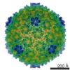 8314MC M: map data used to model this data C: citing same article ( |
|---|---|
| Similar structure data |
-Links
 PDBj
PDBj- Assembly
| Deposited unit | 
|
|---|---|
| 1 | x 60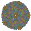
|
| 2 |
|
| 3 | x 5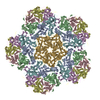
|
| 4 | x 6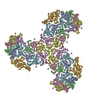
|
| 5 | 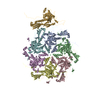
|
| Symmetry | Point symmetry: (Schoenflies symbol: I (icosahedral)) |
-Components
| #1: Protein | Mass: 45590.840 Da / Num. of mol.: 7 / Source method: isolated from a natural source / Source: (natural) Shigella phage Sf6 (virus) / References: UniProt: Q716H0#2: Chemical | ChemComp-CL / |   Mass: 35.453 Da / Num. of mol.: 1 / Source method: obtained synthetically / Formula: Cl Mass: 35.453 Da / Num. of mol.: 1 / Source method: obtained synthetically / Formula: Cl |
|---|
-Experimental details
-Experiment
| Experiment | Method: ELECTRON MICROSCOPY |
|---|---|
| EM experiment | Aggregation state: PARTICLE / 3D reconstruction method: single particle reconstruction |
- Sample preparation
| Component | Name: Shigella phage Sf6 / Type: VIRUS / Entity ID: #1 / Source: NATURAL | |||||||||||||||
|---|---|---|---|---|---|---|---|---|---|---|---|---|---|---|---|---|
| Molecular weight | Value: 19.5 MDa / Experimental value: NO | |||||||||||||||
| Source (natural) | Organism: Shigella phage Sf6 (virus) | |||||||||||||||
| Details of virus | Empty: NO / Enveloped: YES / Isolate: STRAIN / Type: VIRION | |||||||||||||||
| Natural host | Organism: Shigella flexneri / Strain: M94 | |||||||||||||||
| Virus shell | Name: virus capsid / Diameter: 650 nm / Triangulation number (T number): 7 | |||||||||||||||
| Buffer solution | pH: 7.4 | |||||||||||||||
| Buffer component |
| |||||||||||||||
| Specimen | Conc.: 15 mg/ml / Embedding applied: NO / Shadowing applied: NO / Staining applied: NO / Vitrification applied: YES / Details: purified Sf6 phage | |||||||||||||||
| Specimen support | Grid material: COPPER | |||||||||||||||
| Vitrification | Instrument: FEI VITROBOT MARK IV / Cryogen name: ETHANE / Humidity: 100 % / Chamber temperature: 293 K |
- Electron microscopy imaging
| Experimental equipment |  Model: Titan Krios / Image courtesy: FEI Company |
|---|---|
| Microscopy | Model: FEI TITAN KRIOS |
| Electron gun | Electron source:  FIELD EMISSION GUN / Accelerating voltage: 300 kV / Illumination mode: FLOOD BEAM FIELD EMISSION GUN / Accelerating voltage: 300 kV / Illumination mode: FLOOD BEAM |
| Electron lens | Mode: BRIGHT FIELD |
| Image recording | Electron dose: 9 e/Å2 / Detector mode: SUPER-RESOLUTION / Film or detector model: GATAN K2 SUMMIT (4k x 4k) |
- Processing
| EM software |
| ||||||||||||||||||||||||||||
|---|---|---|---|---|---|---|---|---|---|---|---|---|---|---|---|---|---|---|---|---|---|---|---|---|---|---|---|---|---|
| CTF correction | Type: PHASE FLIPPING AND AMPLITUDE CORRECTION | ||||||||||||||||||||||||||||
| 3D reconstruction | Resolution: 2.89 Å / Resolution method: FSC 0.143 CUT-OFF / Num. of particles: 68000 / Symmetry type: POINT | ||||||||||||||||||||||||||||
| Atomic model building | B value: 46.6 / Protocol: AB INITIO MODEL / Space: RECIPROCAL Target criteria: Pseudo-crystallographic R factor and stereochemistry |
