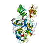
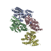
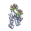
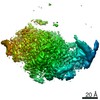
rFVIIIFc is composed of three polypeptide chains. Chain A is the heavy chain of FVIII. Chain B is the light chain of FVIII with a single human IgG1 Fc domain fused to the C terminus of the light chain. Chain C is a free single human IgG1 Fc domain that pairs with the Fc domain at the C terminus of the FVIII light chain. These three chains form the full assembly--a monomeric FVIII with a C-terminal, dimeric Fc-fusion. While all three chains are in the crystal, the dimeric Fc is not visible in the final structure. Silver stain gel of a dissolved crystal confirmed the presence of all three chains.
Mass: 63.546 Da / Num. of mol.: 2 Source method: isolated from a genetically manipulated source Formula: Cu
-
Details
Compound details
Authors state the following: The crystal contains a total of 3 polypeptide chains. Chain A is the ...Authors state the following: The crystal contains a total of 3 polypeptide chains. Chain A is the heavy chain of FVIII. Chain B is the light chain of FVIII fused to a single IgG1 Fc domain. The third chain, which is not indicated here, is another single IgG1 Fc domain that forms a dimer with the single Fc at the C-terminus of the FVIII light chain. There are no coordinates for the either of these Fc halves as there is no density for the dimeric Fc at the C-terminus of FVIII. We have dissolved crystals on a reducing gel and observe the presence of both halves of the Fc (one fused to the C terminus of the FVIII light chain and one free Fc). There are large solvent channels in the crystal that are large enough to accommodate the Fc dimer at the C-terminus of FVIII
Has protein modification
Y
-
Experimental details
-
Experiment
Experiment
Method: X-RAY DIFFRACTION / Number of used crystals: 1
-
Sample preparation
Crystal
Density Matthews: 4.61 Å3/Da / Density % sol: 73.33 %
Crystal grow
Temperature: 297 K / Method: vapor diffusion / pH: 7.3 / Details: 5% Ethanol, 100mM TRIS pH 7.3
Resolution: 4.19→37.6 Å / Cor.coef. Fo:Fc: 0.851 / Cor.coef. Fo:Fc free: 0.715 / SU B: 146.864 / SU ML: 0.828 / Cross valid method: THROUGHOUT / σ(F): 0 / ESU R Free: 0.996 / Stereochemistry target values: MAXIMUM LIKELIHOOD Details: HYDROGENS HAVE BEEN USED IF PRESENT IN THE INPUT U VALUES : WITH TLS ADDED. Authors state the following: The crystal contains a total of 3 polypeptide chains. Chain A is the heavy chain of ...Details: HYDROGENS HAVE BEEN USED IF PRESENT IN THE INPUT U VALUES : WITH TLS ADDED. Authors state the following: The crystal contains a total of 3 polypeptide chains. Chain A is the heavy chain of FVIII. Chain B is the light chain of FVIII fused to a single IgG1 Fc domain. The third chain, which is not indicated here, is another single IgG1 Fc domain that forms a dimer with the single Fc at the C-terminus of the FVIII light chain. There are no coordinates for the either of these Fc halves as there is no density for the dimeric Fc at the C-terminus of FVIII. We have dissolved crystals on a reducing gel and observe the presence of both halves of the Fc (one fused to the C terminus of the FVIII light chain and one free Fc). There are large solvent channels in the crystal that are large enough to accommodate the Fc dimer at the C-terminus of FVIII
Rfactor
Num. reflection
% reflection
Selection details
Rfree
0.3619
1309
5.1 %
RANDOM
Rwork
0.298
-
-
-
obs
0.3011
24550
98.77 %
-
Solvent computation
Ion probe radii: 0.8 Å / Shrinkage radii: 0.8 Å / VDW probe radii: 1.2 Å / Solvent model: MASK
In the structure databanks used in Yorodumi, some data are registered as the other names, "COVID-19 virus" and "2019-nCoV". Here are the details of the virus and the list of structure data.
Jan 31, 2019. EMDB accession codes are about to change! (news from PDBe EMDB page)
EMDB accession codes are about to change! (news from PDBe EMDB page)
The allocation of 4 digits for EMDB accession codes will soon come to an end. Whilst these codes will remain in use, new EMDB accession codes will include an additional digit and will expand incrementally as the available range of codes is exhausted. The current 4-digit format prefixed with “EMD-” (i.e. EMD-XXXX) will advance to a 5-digit format (i.e. EMD-XXXXX), and so on. It is currently estimated that the 4-digit codes will be depleted around Spring 2019, at which point the 5-digit format will come into force.
The EM Navigator/Yorodumi systems omit the EMD- prefix.
Related info.:Q: What is EMD? / ID/Accession-code notation in Yorodumi/EM Navigator
Yorodumi is a browser for structure data from EMDB, PDB, SASBDB, etc.
This page is also the successor to EM Navigator detail page, and also detail information page/front-end page for Omokage search.
The word "yorodu" (or yorozu) is an old Japanese word meaning "ten thousand". "mi" (miru) is to see.
Related info.:EMDB / PDB / SASBDB / Comparison of 3 databanks / Yorodumi Search / Aug 31, 2016. New EM Navigator & Yorodumi / Yorodumi Papers / Jmol/JSmol / Function and homology information / Changes in new EM Navigator and Yorodumi
 Movie
Movie Controller


 Open data
 Basic information
 Components
 Keywords
 Function and homology information
 Homo sapiens (human)
 X-RAY DIFFRACTION /
  Authors
 Citation
 Structure visualization
 Downloads & links
 Other downloads
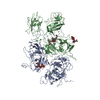


 PDBj
PDBj


 Assembly


 Mass: 40.078 Da / Num. of mol.: 3
 Mass: 63.546 Da / Num. of mol.: 2
 Sample preparation
 / Beamline: 31-ID / Wavelength: 0.9792 Å
 Processing