 Movie
Movie Controller

[English] 日本語
 Yorodumi
- PDB-5jwr: Crystal structure of foldswitch-stabilized KaiB in complex with t... -
+ Open data

- Basic information
| Entry | Database: PDB / ID: 5jwr | |||||||||
|---|---|---|---|---|---|---|---|---|---|---|
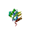
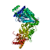
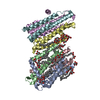
| Title | Crystal structure of foldswitch-stabilized KaiB in complex with the N-terminal CI domain of KaiC and a dimer of KaiA C-terminal domains from Thermosynechococcus elongatus | |||||||||
 Components Components |
| |||||||||
 Keywords Keywords | Transcription Regulator / foldswitch | |||||||||
| Function / homology |  Function and homology information Function and homology informationprotein serine/threonine/tyrosine kinase activity / circadian rhythm / regulation of circadian rhythm / Hydrolases; Acting on acid anhydrides; Acting on acid anhydrides to facilitate cellular and subcellular movement / kinase activity / non-specific serine/threonine protein kinase / protein serine kinase activity / protein serine/threonine kinase activity / regulation of DNA-templated transcription / magnesium ion binding ...protein serine/threonine/tyrosine kinase activity / circadian rhythm / regulation of circadian rhythm / Hydrolases; Acting on acid anhydrides; Acting on acid anhydrides to facilitate cellular and subcellular movement / kinase activity / non-specific serine/threonine protein kinase / protein serine kinase activity / protein serine/threonine kinase activity / regulation of DNA-templated transcription / magnesium ion binding / ATP hydrolysis activity / DNA binding / ATP binding / identical protein binding Similarity search - Function | |||||||||
| Biological species |   Thermosynechococcus elongatus (bacteria) Thermosynechococcus elongatus (bacteria) | |||||||||
| Method |  X-RAY DIFFRACTION / SYNCHROTRON / MOLECULAR REPLACEMENT / Resolution: 2.61 Å X-RAY DIFFRACTION / SYNCHROTRON / MOLECULAR REPLACEMENT / Resolution: 2.61 Å | |||||||||
 Authors Authors | Tseng, R. / Goularte, N.F. / Chavan, A. / Luu, J. / Chang, Y.G. / Heilser, J. / Tripathi, S. / LiWang, A. / Partch, C.L. | |||||||||
| Funding support |  United States, 2items United States, 2items
| |||||||||
 Citation Citation | Journal: Science / Year: 2017 Title: Structural basis of the day-night transition in a bacterial circadian clock. Authors: Tseng, R. / Goularte, N.F. / Chavan, A. / Luu, J. / Cohen, S.E. / Chang, Y.G. / Heisler, J. / Li, S. / Michael, A.K. / Tripathi, S. / Golden, S.S. / LiWang, A. / Partch, C.L. | |||||||||
| History |
|
- Structure visualization
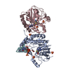
| Structure viewer | Molecule: MolmilJmol/JSmol |
|---|
- Downloads & links
-Download
| PDBx/mmCIF format | 5jwr.cif.gz | 481.8 KB | Display | PDBx/mmCIF format |
|---|---|---|---|---|
| PDB format | pdb5jwr.ent.gz | 399.4 KB | Display | PDB format |
| PDBx/mmJSON format | 5jwr.json.gz | Tree view | PDBx/mmJSON format | |
| Others |  Other downloads Other downloads |
-Validation report
| Arichive directory | https://data.pdbj.org/pub/pdb/validation_reports/jw/5jwrftp://data.pdbj.org/pub/pdb/validation_reports/jw/5jwr | HTTPS FTP |
|---|
-Related structure data


-Links
 PDBj
PDBj

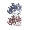
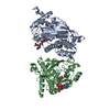
- Assembly
| Deposited unit | 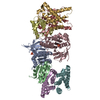
| ||||||||
|---|---|---|---|---|---|---|---|---|---|
| 1 | 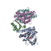
| ||||||||
| 2 | 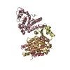
| ||||||||
| Unit cell |
|
-Components
| #1: Protein | Mass: 27738.166 Da / Num. of mol.: 2 / Mutation: R41A, K173A Source method: isolated from a genetically manipulated source Source: (gene. exp.) Thermosynechococcus elongatus (bacteria)Strain: BP-1 / Gene: kaiC, tlr0483 / Plasmid: pET-28b / Production host: References: UniProt: Q79V60, non-specific serine/threonine protein kinase #2: Protein | Mass: 10941.011 Da / Num. of mol.: 2 / Mutation: Y8A, G89A, D91R, Y94A Source method: isolated from a genetically manipulated source Source: (gene. exp.) Thermosynechococcus elongatus (strain BP-1) (bacteria)Strain: BP-1 / Gene: kaiB, tlr0482 / Plasmid: pET-28b / Production host: #3: Protein | Mass: 17332.758 Da / Num. of mol.: 4 / Mutation: C272S Source method: isolated from a genetically manipulated source Source: (gene. exp.) Thermosynechococcus elongatus (bacteria)Strain: BP-1 / Gene: kaiA, tlr0481 / Plasmid: pET-28b / Production host: #4: Chemical |   Mass: 94.971 Da / Num. of mol.: 2 / Source method: obtained synthetically / Formula: PO4 Mass: 94.971 Da / Num. of mol.: 2 / Source method: obtained synthetically / Formula: PO4#5: Water | ChemComp-HOH / |  Mass: 18.015 Da / Num. of mol.: 106 / Source method: isolated from a natural source / Formula: H2O Mass: 18.015 Da / Num. of mol.: 106 / Source method: isolated from a natural source / Formula: H2O |
|---|
-Experimental details
-Experiment
| Experiment | Method: X-RAY DIFFRACTION / Number of used crystals: 1 |
|---|
- Sample preparation
| Crystal | Density Matthews: 3.15 Å3/Da / Density % sol: 60.95 % |
|---|---|
| Crystal grow | Temperature: 295 K / Method: vapor diffusion, hanging drop / pH: 7 Details: 0.15M Ammonium Sulfate, 0.1M Tris pH 7.0, 10% PEG 4000 |
-Data collection
| Diffraction | Mean temperature: 100 K |
|---|---|
| Diffraction source | Source: SYNCHROTRON / Site: ALS / Beamline: 8.3.1 / Wavelength: 1 Å |
| Detector | Type: ADSC QUANTUM 315r / Detector: CCD / Date: Sep 11, 2015 |
| Radiation | Protocol: SINGLE WAVELENGTH / Monochromatic (M) / Laue (L): M / Scattering type: x-ray |
| Radiation wavelength | Wavelength: 1 Å / Relative weight: 1 |
| Reflection | Resolution: 2.6→50 Å / Num. obs: 47380 / % possible obs: 96.4 % / Redundancy: 2 % / Rmerge(I) obs: 0.062 / Net I/σ(I): 21.3 |
| Reflection shell | Resolution: 2.6→2.64 Å / Redundancy: 2.1 % / Rmerge(I) obs: 0.581 / Mean I/σ(I) obs: 2.53 / % possible all: 97.4 |
- Processing
| Software |
| |||||||||||||||||||||||||||||||||||||||||||||||||||||||||||||||||||||||||||||||||||||||||||||||||||||||||||||||||||||||||||||||||||||||||||||||||||||||||||||||||||||||||||||||||||||||||||||||||||||||||||||||||||||||||||||||||
|---|---|---|---|---|---|---|---|---|---|---|---|---|---|---|---|---|---|---|---|---|---|---|---|---|---|---|---|---|---|---|---|---|---|---|---|---|---|---|---|---|---|---|---|---|---|---|---|---|---|---|---|---|---|---|---|---|---|---|---|---|---|---|---|---|---|---|---|---|---|---|---|---|---|---|---|---|---|---|---|---|---|---|---|---|---|---|---|---|---|---|---|---|---|---|---|---|---|---|---|---|---|---|---|---|---|---|---|---|---|---|---|---|---|---|---|---|---|---|---|---|---|---|---|---|---|---|---|---|---|---|---|---|---|---|---|---|---|---|---|---|---|---|---|---|---|---|---|---|---|---|---|---|---|---|---|---|---|---|---|---|---|---|---|---|---|---|---|---|---|---|---|---|---|---|---|---|---|---|---|---|---|---|---|---|---|---|---|---|---|---|---|---|---|---|---|---|---|---|---|---|---|---|---|---|---|---|---|---|---|---|---|---|---|---|---|---|---|---|---|---|---|---|---|---|---|---|
| Refinement | Method to determine structure: MOLECULAR REPLACEMENT / Resolution: 2.61→44.841 Å / SU ML: 0.36 / Cross valid method: FREE R-VALUE / σ(F): 1.96 / Phase error: 27.09
| |||||||||||||||||||||||||||||||||||||||||||||||||||||||||||||||||||||||||||||||||||||||||||||||||||||||||||||||||||||||||||||||||||||||||||||||||||||||||||||||||||||||||||||||||||||||||||||||||||||||||||||||||||||||||||||||||
| Solvent computation | Shrinkage radii: 0.9 Å / VDW probe radii: 1.11 Å | |||||||||||||||||||||||||||||||||||||||||||||||||||||||||||||||||||||||||||||||||||||||||||||||||||||||||||||||||||||||||||||||||||||||||||||||||||||||||||||||||||||||||||||||||||||||||||||||||||||||||||||||||||||||||||||||||
| Refinement step | Cycle: LAST / Resolution: 2.61→44.841 Å
| |||||||||||||||||||||||||||||||||||||||||||||||||||||||||||||||||||||||||||||||||||||||||||||||||||||||||||||||||||||||||||||||||||||||||||||||||||||||||||||||||||||||||||||||||||||||||||||||||||||||||||||||||||||||||||||||||
| Refine LS restraints |
| |||||||||||||||||||||||||||||||||||||||||||||||||||||||||||||||||||||||||||||||||||||||||||||||||||||||||||||||||||||||||||||||||||||||||||||||||||||||||||||||||||||||||||||||||||||||||||||||||||||||||||||||||||||||||||||||||
| LS refinement shell |
| |||||||||||||||||||||||||||||||||||||||||||||||||||||||||||||||||||||||||||||||||||||||||||||||||||||||||||||||||||||||||||||||||||||||||||||||||||||||||||||||||||||||||||||||||||||||||||||||||||||||||||||||||||||||||||||||||
| Refinement TLS params. | Method: refined / Refine-ID: X-RAY DIFFRACTION
| |||||||||||||||||||||||||||||||||||||||||||||||||||||||||||||||||||||||||||||||||||||||||||||||||||||||||||||||||||||||||||||||||||||||||||||||||||||||||||||||||||||||||||||||||||||||||||||||||||||||||||||||||||||||||||||||||
| Refinement TLS group |
|
