Mass: 18.015 Da / Num. of mol.: 144 / Source method: isolated from a natural source / Formula: H2O
-
Details
Sequence details
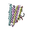
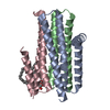
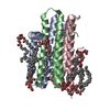
THE PROTEIN CONTAINS AN N-TERMINAL HIS TAG 'GHHHHHHEL'. COMPARED TO THE WILDTYPE FORM, THE PROTEIN ...THE PROTEIN CONTAINS AN N-TERMINAL HIS TAG 'GHHHHHHEL'. COMPARED TO THE WILDTYPE FORM, THE PROTEIN HAS SEVEN MUTATIONS. THEY ARE A41C, C46A, I53V, I70L, M96L, V107D AND C113A.
-
Experimental details
-
Experiment
Experiment
Method: X-RAY DIFFRACTION / Number of used crystals: 2
-
Sample preparation
Crystal
Density Matthews: 3.07 Å3/Da / Density % sol: 59.97 % Description: THE PRESENCE OF ZINC IN THE STRUCTURE WAS CONFIRMED BY A X-RAY FLUORESCENCE SCAN AND WAS SUPPORTED BY A DATA SET COLLECTED AT THE ZINC EDGE (1.282400 A) WHICH IS AVAILABLE IN THIS ENTRY.
Crystal grow
Temperature: 277 K / Method: lipidic cubic phase / pH: 5.6 Details: 3-5%(V/V) 2-METHYL-2, 4-PENTANEDIOL (MPD), 0.1 M SODIUM CHLORIDE, 0.06 M MAGNESIUM ACETATE, 0.1 M SODIUM CITRATE/HCL PH 5.6. CRYSTALLIZED USING THE IN MESO (LIPID CUBIC PHASE) METHOD WITH 7. ...Details: 3-5%(V/V) 2-METHYL-2, 4-PENTANEDIOL (MPD), 0.1 M SODIUM CHLORIDE, 0.06 M MAGNESIUM ACETATE, 0.1 M SODIUM CITRATE/HCL PH 5.6. CRYSTALLIZED USING THE IN MESO (LIPID CUBIC PHASE) METHOD WITH 7.8 MAG AS THE HOST LIPID AT 4 CELSIUS DEGREE.
 Movie
Movie Controller


 Open data
 Basic information
 Components
 Keywords
 Function and homology information
 X-RAY DIFFRACTION /
  Authors
 Citation
 Structure visualization
 Downloads & links
 Other downloads


 PDBj
PDBj


 Assembly
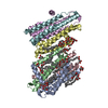

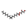
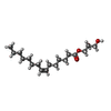


 Mass: 314.460 Da / Num. of mol.: 7 / Source method: obtained synthetically / Formula: C18H34O4
 Mass: 314.460 Da / Num. of mol.: 11 / Source method: obtained synthetically / Formula: C18H34O4
 Mass: 22.990 Da / Num. of mol.: 1 / Source method: obtained synthetically / Formula: Na
 Mass: 189.100 Da / Num. of mol.: 2 / Source method: obtained synthetically / Formula: C6H5O7
 Mass: 65.409 Da / Num. of mol.: 1 / Source method: obtained synthetically / Formula: Zn
 Sample preparation
 / Beamline: 23-ID-B / Wavelength: 1.0332
  Processing