 Movie
Movie Controller


+ Open data

- Basic information
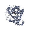
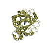
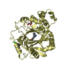
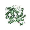
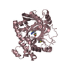
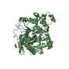
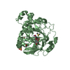
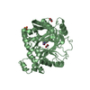
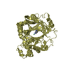
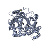
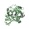
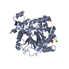
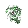
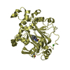
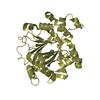
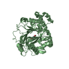
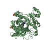
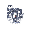
| Entry | Database: PDB / ID: 5a7s | ||||||
|---|---|---|---|---|---|---|---|
| Title | Crystal structure of human JMJD2A in complex with compound 44 | ||||||
 Components Components | LYSINE-SPECIFIC DEMETHYLASE 4A | ||||||
 Keywords Keywords | OXIDOREDUCTASE / JMJD2A / KDM4A | ||||||
| Function / homology |  Function and homology information Function and homology information[histone H3]-trimethyl-L-lysine36 demethylase / histone H3K36me2/H3K36me3 demethylase activity / histone H4K20me2 reader activity / histone H3K36 demethylase activity / cardiac muscle hypertrophy in response to stress / [histone H3]-trimethyl-L-lysine9 demethylase / histone H3K9me2/H3K9me3 demethylase activity / histone H3K9 demethylase activity / histone demethylase activity / pericentric heterochromatin ...[histone H3]-trimethyl-L-lysine36 demethylase / histone H3K36me2/H3K36me3 demethylase activity / histone H4K20me2 reader activity / histone H3K36 demethylase activity / cardiac muscle hypertrophy in response to stress / [histone H3]-trimethyl-L-lysine9 demethylase / histone H3K9me2/H3K9me3 demethylase activity / histone H3K9 demethylase activity / histone demethylase activity / pericentric heterochromatin / NR1H3 & NR1H2 regulate gene expression linked to cholesterol transport and efflux / negative regulation of autophagy / HDMs demethylate histones / fibrillar center / Recruitment and ATM-mediated phosphorylation of repair and signaling proteins at DNA double strand breaks / regulation of gene expression / chromatin remodeling / negative regulation of gene expression / negative regulation of DNA-templated transcription / ubiquitin protein ligase binding / chromatin / zinc ion binding / nucleoplasm / nucleus / cytosol Similarity search - Function | ||||||
| Biological species |  HOMO SAPIENS (human) HOMO SAPIENS (human) | ||||||
| Method |  X-RAY DIFFRACTION / SYNCHROTRON / MOLECULAR REPLACEMENT / Resolution: 2.2 Å X-RAY DIFFRACTION / SYNCHROTRON / MOLECULAR REPLACEMENT / Resolution: 2.2 Å | ||||||
 Authors Authors | Nowak, R. / Velupillai, S. / Krojer, T. / Gileadi, C. / Johansson, C. / Korczynska, M. / Le, D.D. / Younger, N. / Gregori-Puigjane, E. / Tumber, A. ...Nowak, R. / Velupillai, S. / Krojer, T. / Gileadi, C. / Johansson, C. / Korczynska, M. / Le, D.D. / Younger, N. / Gregori-Puigjane, E. / Tumber, A. / Iwasa, E. / Pollock, S.B. / Ortiz Torres, I. / Kopec, J. / Tallant, C. / Froese, S. / von Delft, F. / Arrowsmith, C.H. / Bountra, C. / Edwards, A. / Shoichet, B.K. / Fujimori, D.G. / Oppermann, U. | ||||||
 Citation Citation | Journal: J.Med.Chem. / Year: 2016 Title: Docking and Linking of Fragments to Discover Jumonji Histone Demethylase Inhibitors. Authors: Korczynska, M. / Le, D.D. / Younger, N. / Gregori-Puigjane, E. / Tumber, A. / Krojer, T. / Velupillai, S. / Gileadi, C. / Nowak, R.P. / Iwasa, E. / Pollock, S.B. / Ortiz Torres, I. / ...Authors: Korczynska, M. / Le, D.D. / Younger, N. / Gregori-Puigjane, E. / Tumber, A. / Krojer, T. / Velupillai, S. / Gileadi, C. / Nowak, R.P. / Iwasa, E. / Pollock, S.B. / Ortiz Torres, I. / Oppermann, U. / Shoichet, B.K. / Fujimori, D.G. | ||||||
| History |
|
- Structure visualization
| Structure viewer | Molecule: MolmilJmol/JSmol |
|---|
- Downloads & links
-Download
| PDBx/mmCIF format | 5a7s.cif.gz | 167.7 KB | Display | PDBx/mmCIF format |
|---|---|---|---|---|
| PDB format | pdb5a7s.ent.gz | 131.1 KB | Display | PDB format |
| PDBx/mmJSON format | 5a7s.json.gz | Tree view | PDBx/mmJSON format | |
| Others |  Other downloads Other downloads |
-Validation report
| Arichive directory | https://data.pdbj.org/pub/pdb/validation_reports/a7/5a7sftp://data.pdbj.org/pub/pdb/validation_reports/a7/5a7s | HTTPS FTP |
|---|
-Related structure data
| Related structure data | 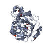 5a7nC 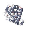 5a7oC 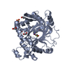 5a7pC 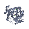 5a7qC 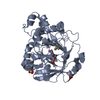 5a7wC 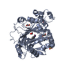 5a80C 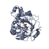 2oq7S C: citing same article ( S: Starting model for refinement |
|---|---|
| Similar structure data |

-Links
 PDBj
PDBj


- Assembly
| Deposited unit | 
| ||||||||
|---|---|---|---|---|---|---|---|---|---|
| 1 | 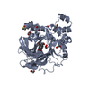
| ||||||||
| 2 | 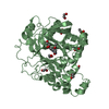
| ||||||||
| Unit cell |
|
-Components
-Protein , 1 types, 2 molecules AB
| #1: Protein | Mass: 44326.273 Da / Num. of mol.: 2 / Fragment: UNP RESIDUES 1-359 Source method: isolated from a genetically manipulated source Source: (gene. exp.) HOMO SAPIENS (human) / Plasmid: PNIC28-BSA4 / Production host:  References: UniProt: O75164, [histone H3]-dimethyl-L-lysine36 demethylase, Oxidoreductases; Acting on paired donors, with incorporation or reduction of molecular oxygen; With 2-oxoglutarate as one ...References: UniProt: O75164, [histone H3]-dimethyl-L-lysine36 demethylase, Oxidoreductases; Acting on paired donors, with incorporation or reduction of molecular oxygen; With 2-oxoglutarate as one donor, and incorporation of one atom of oxygen into each donor |
|---|
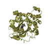
-Non-polymers , 6 types, 417 molecules 


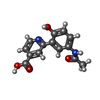


| #2: Chemical |  Mass: 54.938 Da / Num. of mol.: 2 / Source method: obtained synthetically / Formula: Mn Mass: 54.938 Da / Num. of mol.: 2 / Source method: obtained synthetically / Formula: Mn#3: Chemical |  Mass: 65.409 Da / Num. of mol.: 2 / Source method: obtained synthetically / Formula: Zn Mass: 65.409 Da / Num. of mol.: 2 / Source method: obtained synthetically / Formula: Zn#4: Chemical | ChemComp-EDO /  Mass: 62.068 Da / Num. of mol.: 15 / Source method: obtained synthetically / Formula: C2H6O2 Mass: 62.068 Da / Num. of mol.: 15 / Source method: obtained synthetically / Formula: C2H6O2#5: Chemical |  Mass: 272.256 Da / Num. of mol.: 2 / Source method: obtained synthetically / Formula: C14H12N2O4 Mass: 272.256 Da / Num. of mol.: 2 / Source method: obtained synthetically / Formula: C14H12N2O4#6: Chemical |  Mass: 78.133 Da / Num. of mol.: 2 / Source method: obtained synthetically / Formula: C2H6OS / Comment: DMSO, precipitant*YM Mass: 78.133 Da / Num. of mol.: 2 / Source method: obtained synthetically / Formula: C2H6OS / Comment: DMSO, precipitant*YM#7: Water | ChemComp-HOH / | Mass: 18.015 Da / Num. of mol.: 394 / Source method: isolated from a natural source / Formula: H2O |
|---|
-Experimental details
-Experiment
| Experiment | Method: X-RAY DIFFRACTION / Number of used crystals: 1 |
|---|
- Sample preparation
| Crystal | Density Matthews: 2.66 Å3/Da / Density % sol: 2.66 % / Description: NONE |
|---|---|
| Crystal grow | pH: 8.5 Details: 30% PEG3350 -- 0.1M TRIS PH 8.5 -- 0.25M AMMONIUM SULFATE |
-Data collection
| Diffraction | Mean temperature: 100 K |
|---|---|
| Diffraction source | Source: SYNCHROTRON / Site: Diamond  / Beamline: I04 / Wavelength: 0.97964 / Beamline: I04 / Wavelength: 0.97964 |
| Detector | Type: DECTRIS PILATUS 6M / Detector: PIXEL / Date: Mar 7, 2014 |
| Radiation | Protocol: SINGLE WAVELENGTH / Monochromatic (M) / Laue (L): M / Scattering type: x-ray |
| Radiation wavelength | Wavelength: 0.97964 Å / Relative weight: 1 |
| Reflection | Resolution: 2.2→32.76 Å / Num. obs: 43913 / % possible obs: 98.8 % / Observed criterion σ(I): 2.8 / Redundancy: 5.6 % / Rmerge(I) obs: 0.07 / Net I/σ(I): 12.3 |
| Reflection shell | Resolution: 2.2→2.27 Å / Redundancy: 5.3 % / Rmerge(I) obs: 0.56 / Mean I/σ(I) obs: 2.8 / % possible all: 97.8 |
- Processing
| Software |
| ||||||||||||||||
|---|---|---|---|---|---|---|---|---|---|---|---|---|---|---|---|---|---|
| Refinement | Method to determine structure: MOLECULAR REPLACEMENT Starting model: PDB ENTRY 2OQ7 Resolution: 2.2→31.1935 Å / σ(F): 2 / Stereochemistry target values: ML Details: DUAL CONFORMATION MODELED FOR SOME RESIDUES. SOME SIDE CHAINS ARE MISSING. DISORDERED REGIONS HAVE HIGH B FACTORS.
| ||||||||||||||||
| Refinement step | Cycle: LAST / Resolution: 2.2→31.1935 Å
|
