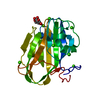
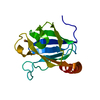
Entry Database : PDB / ID : 5juhTitle Crystal structure of C-terminal domain (RV) of MpAFP Antifreeze protein Keywords / / / Function / homology Function Domain/homology Component
/ / / / / / / Biological species Marinomonas primoryensis (bacteria)Method / / Resolution : 1.35 Å Authors Guo, S. Funding support Organization Grant number Country Canadian Institutes of Health Research (CIHR)
Journal : Sci Adv / Year : 2017Title : Structure of a 1.5-MDa adhesin that binds its Antarctic bacterium to diatoms and ice.Authors: Guo, S. / Stevens, C.A. / Vance, T.D.R. / Olijve, L.L.C. / Graham, L.A. / Campbell, R.L. / Yazdi, S.R. / Escobedo, C. / Bar-Dolev, M. / Yashunsky, V. / Braslavsky, I. / Langelaan, D.N. / ... Authors : Guo, S. / Stevens, C.A. / Vance, T.D.R. / Olijve, L.L.C. / Graham, L.A. / Campbell, R.L. / Yazdi, S.R. / Escobedo, C. / Bar-Dolev, M. / Yashunsky, V. / Braslavsky, I. / Langelaan, D.N. / Smith, S.P. / Allingham, J.S. / Voets, I.K. / Davies, P.L. History Deposition May 10, 2016 Deposition site / Processing site Revision 1.0 Jul 19, 2017 Provider / Type Revision 1.1 Sep 6, 2017 Group / Category / citation_authorItem _citation.country / _citation.journal_abbrev ... _citation.country / _citation.journal_abbrev / _citation.journal_id_CSD / _citation.journal_id_ISSN / _citation.journal_volume / _citation.page_first / _citation.page_last / _citation.pdbx_database_id_DOI / _citation.pdbx_database_id_PubMed / _citation.title / _citation.year Revision 1.2 Sep 20, 2017 Group / Category / Item Revision 1.3 Jan 8, 2020 Group / Category / Item Revision 1.4 Mar 6, 2024 Group / Database references / Derived calculationsCategory chem_comp_atom / chem_comp_bond ... chem_comp_atom / chem_comp_bond / database_2 / pdbx_struct_conn_angle / struct_conn Item _database_2.pdbx_DOI / _database_2.pdbx_database_accession ... _database_2.pdbx_DOI / _database_2.pdbx_database_accession / _pdbx_struct_conn_angle.ptnr1_auth_comp_id / _pdbx_struct_conn_angle.ptnr1_auth_seq_id / _pdbx_struct_conn_angle.ptnr1_label_alt_id / _pdbx_struct_conn_angle.ptnr1_label_asym_id / _pdbx_struct_conn_angle.ptnr1_label_atom_id / _pdbx_struct_conn_angle.ptnr1_label_comp_id / _pdbx_struct_conn_angle.ptnr1_label_seq_id / _pdbx_struct_conn_angle.ptnr1_symmetry / _pdbx_struct_conn_angle.ptnr3_auth_comp_id / _pdbx_struct_conn_angle.ptnr3_auth_seq_id / _pdbx_struct_conn_angle.ptnr3_label_alt_id / _pdbx_struct_conn_angle.ptnr3_label_asym_id / _pdbx_struct_conn_angle.ptnr3_label_atom_id / _pdbx_struct_conn_angle.ptnr3_label_comp_id / _pdbx_struct_conn_angle.ptnr3_label_seq_id / _pdbx_struct_conn_angle.ptnr3_symmetry / _pdbx_struct_conn_angle.value / _struct_conn.pdbx_dist_value / _struct_conn.pdbx_ptnr2_label_alt_id / _struct_conn.ptnr1_auth_comp_id / _struct_conn.ptnr1_auth_seq_id / _struct_conn.ptnr1_label_asym_id / _struct_conn.ptnr1_label_atom_id / _struct_conn.ptnr1_label_comp_id / _struct_conn.ptnr1_label_seq_id / _struct_conn.ptnr2_auth_comp_id / _struct_conn.ptnr2_auth_seq_id / _struct_conn.ptnr2_label_asym_id / _struct_conn.ptnr2_label_atom_id / _struct_conn.ptnr2_label_comp_id / _struct_conn.ptnr2_symmetry
Show all Show less
 Movie
Movie Controller


 Open data
 Basic information
 Components
 Keywords
 Function and homology information
 Marinomonas primoryensis (bacteria)
 X-RAY DIFFRACTION /
  Authors
 Canada, 1items
  Citation
 Structure visualization
 Downloads & links
 Other downloads


 PDBj
PDBj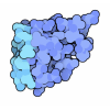
 Assembly
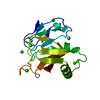

 Mass: 40.078 Da / Num. of mol.: 8 / Source method: obtained synthetically / Formula: Ca
 Mass: 18.015 Da / Num. of mol.: 181 / Source method: isolated from a natural source / Formula: H2O
 Sample preparation
 Processing