 Movie
Movie Controller

[English] 日本語
 Yorodumi
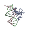
- PDB-3qmc: Structural Basis of Selective Binding of Nonmethylated CpG Island... -
+ Open data

- Basic information
| Entry | Database: PDB / ID: 3qmc | ||||||
|---|---|---|---|---|---|---|---|
| Title | Structural Basis of Selective Binding of Nonmethylated CpG Islands by the CXXC Domain of CFP1 | ||||||
 Components Components |
| ||||||
 Keywords Keywords | DNA BINDING PROTEIN/DNA / Structural Genomics Consortium / SGC / CXXC-type Zn finger / DNA binding / unmethylated CpG motifs / nucleus speckle / DNA BINDING PROTEIN-DNA complex | ||||||
| Function / homology |  Function and homology information Function and homology informationunmethylated CpG binding / XBP1(S) activates chaperone genes / Set1C/COMPASS complex / histone H3K4me3 reader activity / Formation of WDR5-containing histone-modifying complexes / histone methyltransferase complex / cis-regulatory region sequence-specific DNA binding / nuclear matrix / nuclear speck / regulation of DNA-templated transcription ...unmethylated CpG binding / XBP1(S) activates chaperone genes / Set1C/COMPASS complex / histone H3K4me3 reader activity / Formation of WDR5-containing histone-modifying complexes / histone methyltransferase complex / cis-regulatory region sequence-specific DNA binding / nuclear matrix / nuclear speck / regulation of DNA-templated transcription / positive regulation of DNA-templated transcription / nucleoplasm / zinc ion binding / nucleus / cytosol Similarity search - Function | ||||||
| Biological species |  Homo sapiens (human) Homo sapiens (human) | ||||||
| Method |  X-RAY DIFFRACTION / SYNCHROTRON / MOLECULAR REPLACEMENT / molecular replacement / Resolution: 2.1 Å X-RAY DIFFRACTION / SYNCHROTRON / MOLECULAR REPLACEMENT / molecular replacement / Resolution: 2.1 Å | ||||||
 Authors Authors | Lam, R. / Xu, C. / Bian, C.B. / Kania, J. / Bountra, C. / Weigelt, J. / Arrowsmith, C.H. / Edwards, A.M. / Bochkarev, A. / Min, J. / Structural Genomics Consortium (SGC) | ||||||
 Citation Citation | Journal: Nat Commun / Year: 2011 Title: The structural basis for selective binding of non-methylated CpG islands by the CFP1 CXXC domain. Authors: Xu, C. / Bian, C. / Lam, R. / Dong, A. / Min, J. | ||||||
| History |
|
- Structure visualization
| Structure viewer | Molecule: MolmilJmol/JSmol |
|---|
- Downloads & links
-Download
| PDBx/mmCIF format | 3qmc.cif.gz | 66.2 KB | Display | PDBx/mmCIF format |
|---|---|---|---|---|
| PDB format | pdb3qmc.ent.gz | 45.7 KB | Display | PDB format |
| PDBx/mmJSON format | 3qmc.json.gz | Tree view | PDBx/mmJSON format | |
| Others |  Other downloads Other downloads |
-Validation report
| Arichive directory | https://data.pdbj.org/pub/pdb/validation_reports/qm/3qmcftp://data.pdbj.org/pub/pdb/validation_reports/qm/3qmc | HTTPS FTP |
|---|
-Related structure data
| Related structure data |  3qmbSC  3qmdC 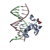 3qmgC 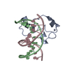 3qmhC  3qmiC S: Starting model for refinement C: citing same article ( |
|---|---|
| Similar structure data |


-Links
 PDBj
PDBj


- Assembly
| Deposited unit | 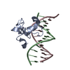
| ||||||||
|---|---|---|---|---|---|---|---|---|---|
| 1 |
| ||||||||
| Unit cell |
|
-Components
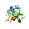
| #1: Protein | Mass: 9527.962 Da / Num. of mol.: 1 / Fragment: CXXC-type Zn finger, residues 161-222 Source method: isolated from a genetically manipulated source Source: (gene. exp.) Homo sapiens (human) / Gene: CXXC1 / Plasmid: pET28-MHL / Production host:  | ||
|---|---|---|---|
| #2: DNA chain | Mass: 3624.356 Da / Num. of mol.: 1 / Fragment: DNA (Nonmethylated CpG Island) / Source method: obtained synthetically Details: DNA was purchased from Integrated DNA Technologies, Inc. | ||
| #3: DNA chain | Mass: 3704.404 Da / Num. of mol.: 1 / Fragment: DNA (Nonmethylated CpG Island) / Source method: obtained synthetically Details: DNA was purchased from Integrated DNA Technologies, Inc. | ||
| #4: Chemical |   Mass: 65.409 Da / Num. of mol.: 2 / Source method: obtained synthetically / Formula: Zn Mass: 65.409 Da / Num. of mol.: 2 / Source method: obtained synthetically / Formula: Zn#5: Water | ChemComp-HOH / |  Mass: 18.015 Da / Num. of mol.: 22 / Source method: isolated from a natural source / Formula: H2O Mass: 18.015 Da / Num. of mol.: 22 / Source method: isolated from a natural source / Formula: H2O |
-Experimental details
-Experiment
| Experiment | Method: X-RAY DIFFRACTION / Number of used crystals: 1 |
|---|
- Sample preparation
| Crystal | Density Matthews: 2.15 Å3/Da / Density % sol: 42.68 % |
|---|---|
| Crystal grow | Temperature: 291 K / Method: vapor diffusion, hanging drop / pH: 7.5 Details: 0.1M HEPES pH 7.5, 0.2M CaCl2, 28% PEG400, VAPOR DIFFUSION, HANGING DROP, temperature 291K |
-Data collection
| Diffraction | Mean temperature: 100 K | ||||||||||||||||||||||||||||||||||||||||||||||||||||||||||||||||||
|---|---|---|---|---|---|---|---|---|---|---|---|---|---|---|---|---|---|---|---|---|---|---|---|---|---|---|---|---|---|---|---|---|---|---|---|---|---|---|---|---|---|---|---|---|---|---|---|---|---|---|---|---|---|---|---|---|---|---|---|---|---|---|---|---|---|---|---|
| Diffraction source | Source: SYNCHROTRON / Site: APS  / Beamline: 19-ID / Wavelength: 0.97924 Å / Beamline: 19-ID / Wavelength: 0.97924 Å | ||||||||||||||||||||||||||||||||||||||||||||||||||||||||||||||||||
| Detector | Type: ADSC QUANTUM 315 / Detector: CCD / Date: Jul 21, 2010 Details: Rosenbaum-Rock high-resolution double-crystal monochromator | ||||||||||||||||||||||||||||||||||||||||||||||||||||||||||||||||||
| Radiation | Monochromator: double-crystal monochromator / Protocol: SINGLE WAVELENGTH / Monochromatic (M) / Laue (L): M / Scattering type: x-ray | ||||||||||||||||||||||||||||||||||||||||||||||||||||||||||||||||||
| Radiation wavelength | Wavelength: 0.97924 Å / Relative weight: 1 | ||||||||||||||||||||||||||||||||||||||||||||||||||||||||||||||||||
| Reflection | Resolution: 2.1→50 Å / Num. obs: 8874 / % possible obs: 100 % / Redundancy: 8.7 % / Rmerge(I) obs: 0.067 / Χ2: 1.089 / Net I/σ(I): 12.9 | ||||||||||||||||||||||||||||||||||||||||||||||||||||||||||||||||||
| Reflection shell |
|
-Phasing
| Phasing | Method: molecular replacement | |||||||||
|---|---|---|---|---|---|---|---|---|---|---|
| Phasing MR | Rfactor: 43.11 / Model details: Phaser MODE: MR_AUTO
|
- Processing
| Software |
| |||||||||||||||||||||||||||||||||||||||||||||||||||||||||||||||||||||||||||||||||||||||||||||||||||||||||||||||||||||||||||||||||||||||||||||||||||
|---|---|---|---|---|---|---|---|---|---|---|---|---|---|---|---|---|---|---|---|---|---|---|---|---|---|---|---|---|---|---|---|---|---|---|---|---|---|---|---|---|---|---|---|---|---|---|---|---|---|---|---|---|---|---|---|---|---|---|---|---|---|---|---|---|---|---|---|---|---|---|---|---|---|---|---|---|---|---|---|---|---|---|---|---|---|---|---|---|---|---|---|---|---|---|---|---|---|---|---|---|---|---|---|---|---|---|---|---|---|---|---|---|---|---|---|---|---|---|---|---|---|---|---|---|---|---|---|---|---|---|---|---|---|---|---|---|---|---|---|---|---|---|---|---|---|---|---|---|
| Refinement | Method to determine structure: MOLECULAR REPLACEMENT Starting model: PDB 3QMB Resolution: 2.1→37.51 Å / Cor.coef. Fo:Fc: 0.95 / Cor.coef. Fo:Fc free: 0.94 / WRfactor Rfree: 0.254 / WRfactor Rwork: 0.22 / Occupancy max: 1 / Occupancy min: 0.4 / SU B: 11.738 / SU ML: 0.139 / Cross valid method: THROUGHOUT / σ(F): 0 / ESU R Free: 0.177 / Stereochemistry target values: MAXIMUM LIKELIHOOD Details: HYDROGENS HAVE BEEN ADDED IN THE RIDING POSITIONS U VALUES : WITH TLS ADDED
| |||||||||||||||||||||||||||||||||||||||||||||||||||||||||||||||||||||||||||||||||||||||||||||||||||||||||||||||||||||||||||||||||||||||||||||||||||
| Solvent computation | Ion probe radii: 0.8 Å / Shrinkage radii: 0.8 Å / VDW probe radii: 1.4 Å / Solvent model: BABINET MODEL WITH MASK | |||||||||||||||||||||||||||||||||||||||||||||||||||||||||||||||||||||||||||||||||||||||||||||||||||||||||||||||||||||||||||||||||||||||||||||||||||
| Displacement parameters | Biso max: 125.73 Å2 / Biso mean: 53.3165 Å2 / Biso min: 26.5 Å2
| |||||||||||||||||||||||||||||||||||||||||||||||||||||||||||||||||||||||||||||||||||||||||||||||||||||||||||||||||||||||||||||||||||||||||||||||||||
| Refinement step | Cycle: LAST / Resolution: 2.1→37.51 Å
| |||||||||||||||||||||||||||||||||||||||||||||||||||||||||||||||||||||||||||||||||||||||||||||||||||||||||||||||||||||||||||||||||||||||||||||||||||
| Refine LS restraints |
| |||||||||||||||||||||||||||||||||||||||||||||||||||||||||||||||||||||||||||||||||||||||||||||||||||||||||||||||||||||||||||||||||||||||||||||||||||
| LS refinement shell | Refine-ID: X-RAY DIFFRACTION / Total num. of bins used: 20
| |||||||||||||||||||||||||||||||||||||||||||||||||||||||||||||||||||||||||||||||||||||||||||||||||||||||||||||||||||||||||||||||||||||||||||||||||||
| Refinement TLS params. | Method: refined / Refine-ID: X-RAY DIFFRACTION
| |||||||||||||||||||||||||||||||||||||||||||||||||||||||||||||||||||||||||||||||||||||||||||||||||||||||||||||||||||||||||||||||||||||||||||||||||||
| Refinement TLS group |
|
