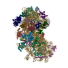
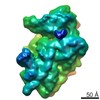
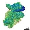
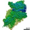
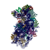
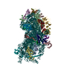
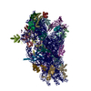
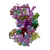
- PDB-5it9: Structure of the yeast Kluyveromyces lactis small ribosomal subun... -
+
データを開く
IDまたはキーワード:
読み込み中...
-
基本情報
登録情報
データベース: PDB / ID: 5it9
タイトル
Structure of the yeast Kluyveromyces lactis small ribosomal subunit in complex with the cricket paralysis virus IRES.
要素
(Ribosomal protein ...) x 33
18S ribosomal RNA
Cricket paralysis virus IRES RNA
キーワード
RIBOSOME / IRES / small / subunit
機能・相同性
機能・相同性情報
90S preribosome / translation regulator activity / maturation of SSU-rRNA from tricistronic rRNA transcript (SSU-rRNA, 5.8S rRNA, LSU-rRNA) / maturation of SSU-rRNA / small-subunit processome / rRNA processing / ribosome binding / ribosomal small subunit biogenesis / ribosomal small subunit assembly / small ribosomal subunit ...90S preribosome / translation regulator activity / maturation of SSU-rRNA from tricistronic rRNA transcript (SSU-rRNA, 5.8S rRNA, LSU-rRNA) / maturation of SSU-rRNA / small-subunit processome / rRNA processing / ribosome binding / ribosomal small subunit biogenesis / ribosomal small subunit assembly / small ribosomal subunit / small ribosomal subunit rRNA binding / cytosolic small ribosomal subunit / cytoplasmic translation / rRNA binding / structural constituent of ribosome / ribosome / translation / ribonucleoprotein complex / mRNA binding / nucleolus / RNA binding / zinc ion binding / nucleus / cytosol / cytoplasm 類似検索 - 分子機能
N-terminal domain of TfIIb - #150 / Single alpha-helices involved in coiled-coils or other helix-helix interfaces - #2650 / Ribosomal protein S26 / Ribosomal protein S8e, subdomain / Ribosomal protein S17 / Ribosomal protein S4, central domain / Phosducin; domain 2 / OB fold (Dihydrolipoamide Acetyltransferase, E2P) - #1000 / Ribosomal protein S21 / Hypothetical Cytosolic Protein; Chain: A; ...N-terminal domain of TfIIb - #150 / Single alpha-helices involved in coiled-coils or other helix-helix interfaces - #2650 / Ribosomal protein S26 / Ribosomal protein S8e, subdomain / Ribosomal protein S17 / Ribosomal protein S4, central domain / Phosducin; domain 2 / OB fold (Dihydrolipoamide Acetyltransferase, E2P) - #1000 / Ribosomal protein S21 / Hypothetical Cytosolic Protein; Chain: A; / Ribosomal protein S27 / first zn-finger domain of poly(adp-ribose) polymerase-1 / Alpha-Beta Plaits - #3370 / Diphtheria Toxin Repressor; domain 2 / N-terminal domain of TfIIb / Ribosomal protein S3, C-terminal domain / Ribosomal Protein S14/S29 / 30s Ribosomal Protein S14; Chain N / Ribosomal Protein S8; Chain: A, domain 1 - #30 / Ribosomal protein S3 C-terminal domain / Dna Ligase; domain 1 - #10 / Ribosomal protein S11/S14 / Ribosomal protein S10 / S15/NS1, RNA-binding / Ribosomal Protein S7 / Ribosomal protein S7/S5 / RNA-binding S4 domain / SH3 type barrels. - #30 / Ribosomal protein L30/S12 / Structural Genomics Hypothetical 15.5 Kd Protein In mrcA-pckA Intergenic Region; Chain A / K homology (KH) domain / Helicase, Ruva Protein; domain 3 - #50 / Double Stranded RNA Binding Domain - #20 / Glucose-6-phosphate isomerase like protein; domain 1 / Ribosomal Protein S8; Chain: A, domain 1 / Other non-globular / N-terminal domain of TfIIb / Ribosomal Protein S5; domain 2 - #10 / GMP Synthetase; Chain A, domain 3 / Ribosomal Protein S5; domain 2 / Double Stranded RNA Binding Domain / : / Ribosomal protein S26e signature. / Ribosomal protein S21e, conserved site / Ribosomal protein S21e signature. / : / Ribosomal protein S12e signature. / Ribosomal protein S26e / Ribosomal protein S26e superfamily / Ribosomal protein S26e / Ribosomal protein S12e / 60s Ribosomal Protein L30; Chain: A; / Ribosomal protein S19e, conserved site / Ribosomal protein S19e signature. / Ribosomal protein S5, eukaryotic/archaeal / Small (40S) ribosomal subunit Asc1/RACK1 / Ribosomal protein S21e / Ribosomal protein S21e superfamily / Ribosomal protein S21e / Ribosomal protein S2, eukaryotic / S27a-like superfamily / : / Ribosomal protein S7e signature. / 40S Ribosomal protein S10 / Plectin/S10, N-terminal / Plectin/S10 domain / Ribosomal protein S8e subdomain, eukaryotes / Ribosomal protein S10, eukaryotic/archaeal / Ribosomal protein S3Ae, conserved site / Ribosomal protein S3Ae signature. / Ribosomal protein S30 / Ribosomal protein S17e, conserved site / Ribosomal protein S30 / Ribosomal protein S17e signature. / Ribosomal protein S27a / Ribosomal protein S25 / Ribosomal protein S27a / S25 ribosomal protein / Ribosomal protein S27a / Ribosomal protein S2, eukaryotic/archaeal / Ribosomal protein S27e signature. / 40S ribosomal protein S29/30S ribosomal protein S14 type Z / Ribosomal protein S4e, N-terminal, conserved site / Ribosomal protein S4e signature. / 40S ribosomal protein S4, C-terminal domain / 40S ribosomal protein S4 C-terminus / Ribosomal protein S8e, conserved site / Ribosomal protein S8e signature. / Ribosomal protein S3, eukaryotic/archaeal / Ribosomal protein S19e / Ribosomal protein S19e / Ribosomal_S19e / Ribosomal protein S6, eukaryotic / Single Sheet / Ribosomal protein S7e / Ribosomal protein S7e / 40S ribosomal protein S1/3, eukaryotes / Ribosomal protein S19A/S15e / 40S ribosomal protein S11, N-terminal / Ribosomal_S17 N-terminal 類似検索 - ドメイン・相同性
: / RNA / RNA (> 10) / RNA (> 100) / RNA (> 1000) / Small ribosomal subunit protein uS11 / Small ribosomal subunit protein eS28 / Ubiquitin-ribosomal protein eS31 fusion protein / Small ribosomal subunit protein uS10 / KLLA0F18040p ...: / RNA / RNA (> 10) / RNA (> 100) / RNA (> 1000) / Small ribosomal subunit protein uS11 / Small ribosomal subunit protein eS28 / Ubiquitin-ribosomal protein eS31 fusion protein / Small ribosomal subunit protein uS10 / KLLA0F18040p / Small ribosomal subunit protein uS5 / KLLA0F07843p / 40S ribosomal protein S12 / Small ribosomal subunit protein eS6 / RP21 / 40S ribosomal protein S8 / Small ribosomal subunit protein uS2 / Small ribosomal subunit protein RACK1 / 40S ribosomal protein S27 / Small ribosomal subunit protein uS14 / KLLA0D10659p / Small ribosomal subunit protein uS3 / 40S ribosomal protein S26 / 40S ribosomal protein S7 / 40S ribosomal protein S24 / 40S ribosomal protein S30 / KLLA0B08173p / Small ribosomal subunit protein uS8 / 40S ribosomal protein S25 / Small ribosomal subunit protein eS1 / 40S ribosomal protein S4 / KLLA0B01562p / KLLA0B01474p / RP41 / S16a / Small ribosomal subunit protein eS21 / RPS23 / Small ribosomal subunit protein uS9 類似検索 - 構成要素
ジャーナル: Elife / 年: 2016 タイトル: Structural characterization of ribosome recruitment and translocation by type IV IRES. 著者: Jason Murray / Christos G Savva / Byung-Sik Shin / Thomas E Dever / V Ramakrishnan / Israel S Fernández / 要旨: Viral mRNA sequences with a type IV IRES are able to initiate translation without any host initiation factors. Initial recruitment of the small ribosomal subunit as well as two translocation steps ...Viral mRNA sequences with a type IV IRES are able to initiate translation without any host initiation factors. Initial recruitment of the small ribosomal subunit as well as two translocation steps before the first peptidyl transfer are essential for the initiation of translation by these mRNAs. Using electron cryomicroscopy (cryo-EM) we have structurally characterized at high resolution how the Cricket Paralysis Virus Internal Ribosomal Entry Site (CrPV-IRES) binds the small ribosomal subunit (40S) and the translocation intermediate stabilized by elongation factor 2 (eEF2). The CrPV-IRES restricts tvhe otherwise flexible 40S head to a conformation compatible with binding the large ribosomal subunit (60S). Once the 60S is recruited, the binary CrPV-IRES/80S complex oscillates between canonical and rotated states (Fernández et al., 2014; Koh et al., 2014), as seen for pre-translocation complexes with tRNAs. Elongation factor eEF2 with a GTP analog stabilizes the ribosome-IRES complex in a rotated state with an extra ~3 degrees of rotation. Key residues in domain IV of eEF2 interact with pseudoknot I (PKI) of the CrPV-IRES stabilizing it in a conformation reminiscent of a hybrid tRNA state. The structure explains how diphthamide, a eukaryotic and archaeal specific post-translational modification of a histidine residue of eEF2, is involved in translocation.
#89 - 2007年5月 アコニターゼと鉄調節タンパク質1 (Aconitase and Iron Regulatory Protein 1) 類似性 (1)
#241 - 2020年1月 20年の分子を振り返って (Twenty Years of Molecules) 類似性 (1)
-
集合体
登録構造単位
A: Ribosomal protein uS2 B: Ribosomal protein eS1 C: Ribosomal protein uS5 D: Ribosomal protein uS3 E: Ribosomal protein eS4 F: Ribosomal protein uS7 G: Ribosomal protein eS6 H: Ribosomal protein eS7 I: Ribosomal protein eS8 J: Ribosomal protein uS4 K: Ribosomal protein eS10 L: Ribosomal protein uS17 M: Ribosomal protein eS12 N: Ribosomal protein uS15 O: Ribosomal protein uS14 P: Ribosomal protein uS19 Q: Ribosomal protein uS9 R: Ribosomal protein eS17 S: Ribosomal protein uS13 T: Ribosomal protein eS19 U: Ribosomal protein uS10 V: Ribosomal protein eS21 W: Ribosomal protein uS8 X: Ribosomal protein uS21 Y: Ribosomal protein eS24 Z: Ribosomal protein eS25 a: Ribosomal protein eS26 b: Ribosomal protein eS27 c: Ribosomal protein eS28 d: Ribosomal protein eS29 e: Ribosomal protein eS30 f: Ribosomal protein eS31 g: Ribosomal protein RACK1 2: 18S ribosomal RNA i: Cricket paralysis virus IRES RNA ヘテロ分子
装置: FEI VITROBOT MARK II / 凍結剤: ETHANE / 湿度: 100 % / 凍結前の試料温度: 298 K
-
電子顕微鏡撮影
実験機器
モデル: Titan Krios / 画像提供: FEI Company
顕微鏡
モデル: FEI TITAN KRIOS
電子銃
電子線源: FIELD EMISSION GUN / 加速電圧: 300 kV / 照射モード: OTHER
電子レンズ
モード: BRIGHT FIELD
試料ホルダ
試料ホルダーモデル: FEI TITAN KRIOS AUTOGRID HOLDER
撮影
電子線照射量: 25 e/Å2 / 検出モード: INTEGRATING フィルム・検出器のモデル: FEI FALCON II (4k x 4k)
-
解析
ソフトウェア
名称: REFMAC / バージョン: 5.8.0124 / 分類: 精密化
EMソフトウェア
ID
名称
バージョン
カテゴリ
1
EPU
粒子像選択
2
EPU
1.8
画像取得
4
CTFFIND
CTF補正
9
RELION
1.4
初期オイラー角割当
13
REFMAC
5.8
モデル精密化
CTF補正
タイプ: PHASE FLIPPING ONLY
3次元再構成
解像度: 3.8 Å / 解像度の算出法: FSC 0.143 CUT-OFF / 粒子像の数: 54481 / アルゴリズム: BACK PROJECTION / 対称性のタイプ: POINT
原子モデル構築
プロトコル: OTHER / 空間: RECIPROCAL
精密化
解像度: 3.8→257.28 Å / Cor.coef. Fo:Fc: 0.848 / SU B: 43.619 / SU ML: 0.614 / σ(F): 0 / ESU R: 0.945 立体化学のターゲット値: MAXIMUM LIKELIHOOD WITH PHASES 詳細: HYDROGENS HAVE BEEN ADDED IN THE RIDING POSITIONS U VALUES : REFINED INDIVIDUALLY
 ムービー
ムービー コントローラー


 データを開く
 基本情報
基本情報 要素
要素 キーワード
キーワード 機能・相同性情報
 Cricket paralysis virus (ウイルス)
 Kluyveromyces lactis (酵母)
 データ登録者
 英国,
英国,  米国, 3件
  引用
引用 構造の表示
構造の表示 ダウンロードとリンク
 その他のダウンロード
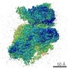


 PDBj
PDBj


 集合体
集合体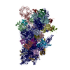


 分子量: 24.305 Da / 分子数: 80 / 由来タイプ: 合成 / 式: Mg
 分子量: 65.409 Da / 分子数: 3 / 由来タイプ: 合成 / 式: Zn
 試料調製
試料調製 電子顕微鏡撮影
 FIELD EMISSION GUN / 加速電圧: 300 kV / 照射モード: OTHER
 解析
解析