 Movie
Movie Controller

[English] 日本語
 Yorodumi
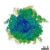
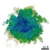
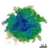
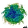
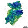
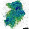
- EMDB-20249: Structure of a mammalian small ribosomal subunit in complex with ... -
+ Open data

- Basic information
| Entry | Database: EMDB / ID: EMD-20249 | |||||||||
|---|---|---|---|---|---|---|---|---|---|---|
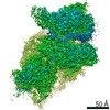
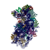
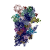
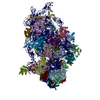
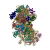
| Title | Structure of a mammalian small ribosomal subunit in complex with the Israeli Acute Paralysis Virus IRES (Class 2) | |||||||||
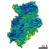
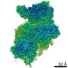
 Map data Map data | ||||||||||
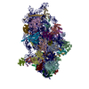
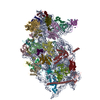
 Sample Sample |
| |||||||||
 Keywords Keywords | Israeli Acute Paralysis Virus / Internal Ribosome Entry Site / IRES / Small Ribosomal Subunit / 40S / RIBOSOME | |||||||||
| Function / homology |  Function and homology information Function and homology informationlaminin receptor activity / ubiquitin ligase inhibitor activity / 90S preribosome / positive regulation of signal transduction by p53 class mediator / phagocytic cup / translation regulator activity / rough endoplasmic reticulum / laminin binding / ribosomal small subunit export from nucleus / gastrulation ...laminin receptor activity / ubiquitin ligase inhibitor activity / 90S preribosome / positive regulation of signal transduction by p53 class mediator / phagocytic cup / translation regulator activity / rough endoplasmic reticulum / laminin binding / ribosomal small subunit export from nucleus / gastrulation / MDM2/MDM4 family protein binding / class I DNA-(apurinic or apyrimidinic site) endonuclease activity / cytosolic ribosome / DNA-(apurinic or apyrimidinic site) lyase / maturation of SSU-rRNA from tricistronic rRNA transcript (SSU-rRNA, 5.8S rRNA, LSU-rRNA) / positive regulation of apoptotic signaling pathway / maturation of SSU-rRNA / small-subunit processome / spindle / rRNA processing / rhythmic process / positive regulation of canonical Wnt signaling pathway / regulation of translation / ribosomal small subunit assembly / virus receptor activity / ribosome binding / ribosomal small subunit biogenesis / small ribosomal subunit / small ribosomal subunit rRNA binding / cytosolic small ribosomal subunit / perikaryon / cell differentiation / cytoplasmic translation / mitochondrial inner membrane / postsynaptic density / rRNA binding / structural constituent of ribosome / ribosome / translation / ribonucleoprotein complex / cell division / DNA repair / mRNA binding / apoptotic process / centrosome / synapse / dendrite / nucleolus / perinuclear region of cytoplasm / Golgi apparatus / DNA binding / RNA binding / zinc ion binding / membrane / nucleus / plasma membrane / cytoplasm Similarity search - Function | |||||||||
| Biological species |   Israeli acute paralysis virus Israeli acute paralysis virus | |||||||||
| Method | single particle reconstruction / cryo EM / Resolution: 3.2 Å | |||||||||
 Authors Authors | Acosta-Reyes FJ / Neupane R | |||||||||
| Funding support |  United States, 1 items United States, 1 items
| |||||||||
 Citation Citation | Journal: EMBO J / Year: 2019 Title: The Israeli acute paralysis virus IRES captures host ribosomes by mimicking a ribosomal state with hybrid tRNAs. Authors: Francisco Acosta-Reyes / Ritam Neupane / Joachim Frank / Israel S Fernández / Abstract: Colony collapse disorder (CCD) is a multi-faceted syndrome decimating bee populations worldwide, and a group of viruses of the widely distributed Dicistroviridae family have been identified as a ...Colony collapse disorder (CCD) is a multi-faceted syndrome decimating bee populations worldwide, and a group of viruses of the widely distributed Dicistroviridae family have been identified as a causing agent of CCD. This family of viruses employs non-coding RNA sequences, called internal ribosomal entry sites (IRESs), to precisely exploit the host machinery for viral protein production. Using single-particle cryo-electron microscopy (cryo-EM), we have characterized how the IRES of Israeli acute paralysis virus (IAPV) intergenic region captures and redirects translating ribosomes toward viral RNA messages. We reconstituted two in vitro reactions targeting a pre-translocation and a post-translocation state of the IAPV-IRES in the ribosome, allowing us to identify six structures using image processing classification methods. From these, we reconstructed the trajectory of IAPV-IRES from the early small subunit recruitment to the final post-translocated state in the ribosome. An early commitment of IRES/ribosome complexes for global pre-translocation mimicry explains the high efficiency observed for this IRES. Efforts directed toward fighting CCD by targeting the IAPV-IRES using RNA-interference technology are underway, and the structural framework presented here may assist in further refining these approaches. | |||||||||
| History |
|
- Structure visualization
| Movie |
Movie viewer |
|---|---|
| Structure viewer | EM map: SurfViewMolmilJmol/JSmol |
| Supplemental images |

- Downloads & links
-EMDB archive
| Map data | emd_20249.map.gz | 12.8 MB | EMDB map data format | |
|---|---|---|---|---|
| Header (meta data) | emd-20249-v30.xmlemd-20249.xml | 58.1 KB 58.1 KB | Display Display | EMDB header |
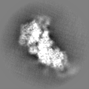
| Images | 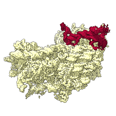 emd_20249.png emd_20249.png | 172.5 KB | ||
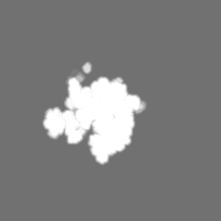
| Masks | emd_20249_msk_1.map | 115.9 MB | Mask map | |
| Filedesc metadata | emd-20249.cif.gz | 12.2 KB | ||
| Others | emd_20249_additional.map.gzemd_20249_half_map_1.map.gzemd_20249_half_map_2.map.gz | 90.8 MB 91.1 MB 91.1 MB | ||
| Archive directory |  http://ftp.pdbj.org/pub/emdb/structures/EMD-20249ftp://ftp.pdbj.org/pub/emdb/structures/EMD-20249 http://ftp.pdbj.org/pub/emdb/structures/EMD-20249ftp://ftp.pdbj.org/pub/emdb/structures/EMD-20249 | HTTPS FTP |
-Related structure data
| Related structure data |  6p4hMC  6p4gC  6p5iC  6p5jC  6p5kC  6p5nC C: citing same article ( M: atomic model generated by this map |
|---|---|
| Similar structure data |
-Links
| EMDB pages | EMDB (EBI/PDBe) / EMDataResource |
|---|---|
| Related items in Molecule of the Month |


-Map
| File | Download / File: emd_20249.map.gz / Format: CCP4 / Size: 115.9 MB / Type: IMAGE STORED AS FLOATING POINT NUMBER (4 BYTES) | ||||||||||||||||||||||||||||||||||||||||||||||||||||||||||||
|---|---|---|---|---|---|---|---|---|---|---|---|---|---|---|---|---|---|---|---|---|---|---|---|---|---|---|---|---|---|---|---|---|---|---|---|---|---|---|---|---|---|---|---|---|---|---|---|---|---|---|---|---|---|---|---|---|---|---|---|---|---|
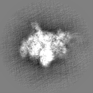
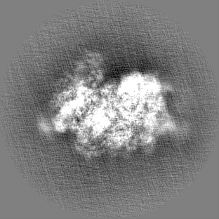
| Projections & slices | Image control
Images are generated by Spider. | ||||||||||||||||||||||||||||||||||||||||||||||||||||||||||||
| Voxel size | X=Y=Z: 1.233 Å | ||||||||||||||||||||||||||||||||||||||||||||||||||||||||||||
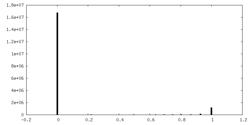
| Density |
| ||||||||||||||||||||||||||||||||||||||||||||||||||||||||||||
| Symmetry | Space group: 1 | ||||||||||||||||||||||||||||||||||||||||||||||||||||||||||||
| Details | EMDB XML:
CCP4 map header:
| ||||||||||||||||||||||||||||||||||||||||||||||||||||||||||||
 Z (Sec.)
 Y (Row.)
 X (Col.)


-Supplemental data
-Mask #1
| File | emd_20249_msk_1.map | ||||||||||||
|---|---|---|---|---|---|---|---|---|---|---|---|---|---|
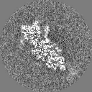
| Projections & Slices |
| ||||||||||||
| Density Histograms |


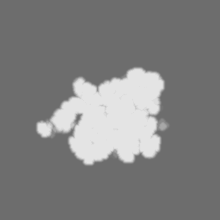

-Additional map: #1
| File | emd_20249_additional.map | ||||||||||||
|---|---|---|---|---|---|---|---|---|---|---|---|---|---|
| Projections & Slices |
| ||||||||||||
| Density Histograms |


-Half map: #1
| File | emd_20249_half_map_1.map | ||||||||||||
|---|---|---|---|---|---|---|---|---|---|---|---|---|---|
| Projections & Slices |
| ||||||||||||
| Density Histograms |


-Half map: #2
| File | emd_20249_half_map_2.map | ||||||||||||
|---|---|---|---|---|---|---|---|---|---|---|---|---|---|
| Projections & Slices |
| ||||||||||||
| Density Histograms |


- Sample components
+Entire : Structure of a mammalian small ribosomal subunit in complex with ...
+Supramolecule #1: Structure of a mammalian small ribosomal subunit in complex with ...
+Macromolecule #1: 18S rRNA
+Macromolecule #6: IAPV-IRES
+Macromolecule #2: uS2
+Macromolecule #3: eS1
+Macromolecule #4: uS5
+Macromolecule #5: uS3
+Macromolecule #7: eS4
+Macromolecule #8: uS7
+Macromolecule #9: eS6
+Macromolecule #10: eS7
+Macromolecule #11: eS8
+Macromolecule #12: uS4
+Macromolecule #13: eS10
+Macromolecule #14: uS17
+Macromolecule #15: eS12
+Macromolecule #16: uS15
+Macromolecule #17: uS11
+Macromolecule #18: uS19
+Macromolecule #19: uS9
+Macromolecule #20: eS17
+Macromolecule #21: uS13
+Macromolecule #22: eS19
+Macromolecule #23: uS10
+Macromolecule #24: eS21
+Macromolecule #25: uS8
+Macromolecule #26: uS12
+Macromolecule #27: eS24
+Macromolecule #28: eS25
+Macromolecule #29: eS26
+Macromolecule #30: eS27
+Macromolecule #31: eS28
+Macromolecule #32: eS29
+Macromolecule #33: eS30
+Macromolecule #34: eS31
+Macromolecule #35: RACK1
+Macromolecule #36: eL41
-Experimental details
-Structure determination
| Method | cryo EM |
|---|---|
 Processing Processing | single particle reconstruction |
| Aggregation state | particle |
-Sample preparation #1
| Preparation ID | 1 | ||||||||||
|---|---|---|---|---|---|---|---|---|---|---|---|
| Buffer | pH: 7.5 Component:
| ||||||||||
| Grid | Model: Quantifoil R2/2 / Material: COPPER / Support film - Film type ID: 1 / Support film - Material: CARBON / Support film - topology: HOLEY / Support film - Film thickness: 5 / Pretreatment - Type: PLASMA CLEANING / Pretreatment - Time: 25 sec. / Pretreatment - Atmosphere: OTHER / Pretreatment - Pressure: 9.33257 kPa Details: Plasma cleaning for both holey carbon and holey gold grids was done on a Gatan Solarus with Hydrogen (6.4 sccm gas flow) and Oxygen (27.5 sccm gas flow) and 10 W cleaning power. | ||||||||||
| Vitrification | Cryogen name: ETHANE / Chamber humidity: 100 % / Chamber temperature: 277.15 K / Instrument: FEI VITROBOT MARK IV | ||||||||||
| Details | Ribosomal complexes for the pre-translocated state were assembled at 240-390 nM concentration and applied to plasma treated holey carbon. |
-Sample preparation #2
| Preparation ID | 2 | ||||||||||
|---|---|---|---|---|---|---|---|---|---|---|---|
| Buffer | pH: 7.5 Component:
| ||||||||||
| Grid | Model: Quantifoil R2/2 / Material: COPPER / Support film - Film type ID: 1 / Support film - Material: CARBON / Support film - topology: HOLEY / Support film - Film thickness: 5 / Pretreatment - Type: PLASMA CLEANING / Pretreatment - Time: 25 sec. / Pretreatment - Atmosphere: OTHER / Pretreatment - Pressure: 9.33257 kPa Details: Plasma cleaning for both holey carbon and holey gold grids was done on a Gatan Solarus with Hydrogen (6.4 sccm gas flow) and Oxygen (27.5 sccm gas flow) and 10 W cleaning power. | ||||||||||
| Vitrification | Cryogen name: ETHANE / Chamber humidity: 100 % / Chamber temperature: 277.15 K / Instrument: FEI VITROBOT MARK IV | ||||||||||
| Details | Same sample and buffer conditions used for the holey carbon grids was used for the holey gold grids. |
- Electron microscopy
| Microscope | FEI TECNAI F30 |
|---|---|
| Image recording | Film or detector model: GATAN K2 SUMMIT (4k x 4k) / Detector mode: COUNTING / Digitization - Dimensions - Width: 3710 pixel / Digitization - Dimensions - Height: 3838 pixel / Digitization - Frames/image: 1-40 / Number real images: 11234 / Average exposure time: 8.0 sec. / Average electron dose: 42.09 e/Å2 |
| Electron beam | Acceleration voltage: 300 kV / Electron source:  FIELD EMISSION GUN FIELD EMISSION GUN |
| Electron optics | Illumination mode: OTHER / Imaging mode: BRIGHT FIELD / Cs: 2.26 mm / Nominal defocus max: 2.5 µm / Nominal defocus min: 0.8 µm / Nominal magnification: 31000 |
| Sample stage | Cooling holder cryogen: NITROGEN |
| Experimental equipment |  Model: Tecnai F30 / Image courtesy: FEI Company |

